Cardiac MRI > Pathology > Cardiomyopathies > Myocarditis
Myocarditis
![]()
The most common cause of myocarditis is viral infection, usually with Coxsackievirus B. Inflamed myocardium hyperenhances on early gadolinium enhanced T1 weighted images due to increased inflow of blood. Delayed enhanced imaging will demonstrate enhancement in the mid-myocardium, often in a patchy pattern. T2 weighted images will be bright due to edema.
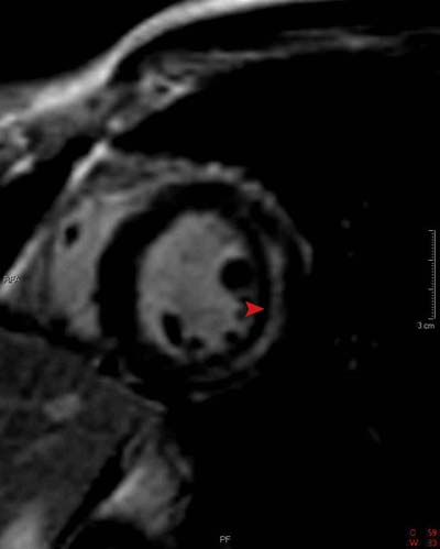
This IR image from a patient with myocarditis shows delayed hyperenhancement (arrowhead). However, unlike the delayed hyperenhancement seen in MI, this image shows enhancement only in the mid-myocardial wall and none in the subendocardium. (Note: there is an area of black immediately between the blood pool and area of enhancement that represents normal subendocardium).
![]()
![]()