Cardiac MRI > Pathology > Pericardial Disease > Pericardial Effusion
Pericardial Effusion
![]()
Pericardial effusion is fluid in the pericardial space. Patients can present with pain, dyspnea, pericardial friction rub, and hemodynamic compromise. Common causes include neoplasm, uremia, autoimmune disease, inflammation, viral infection, tuberculosis, and hemopericardium. A small amount of fluid is relatively inconsequential, but large or rapidly accumulating effusions can cause tamponade.
Cardiac MRI can be used to characterize pericardial effusions and assess the pericardium. Major indications for cardiac MRI is to evaluate for loculated pericardial effusions and pericardial adhessions, determine if an effusion is complicatated (hemorrhagic vs. non-hemorrhagic), measure pericardial thickness, and assess for pericardial enhancement. On spin-echo imaging, simple effusions have low signal intensity on T1-weighted imaging and a high signal intensity on T2-weighted imaging. Chylous and hemorrhagic effusions have a higher signal on T1-weighted imaging. Cardiac MRI is also useful in patients with inconclusive echocardiographic exams.
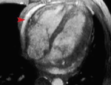
This two chamber long axis gradient echo image shows a bright fluid collection in the pericardial space (arrowhead). If this fluid accumulated quickly, the patient may experience symptoms of cardiac tamponade.
![]()
![]()