Cardiac MRI > Post Test

Post-Test

All of the material covered in this test can be found on the previous pages. Answers are provided at the end of the test.
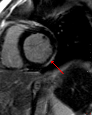
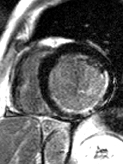
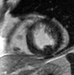
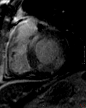
1) This gradient echo cine shows which of the following abnormalities?


Ventricular septal defect
Right ventricular hypertrophy
Aortic regurgitation and stenosis
Aortic regurgitation
Aortic stenosis


2) This inversion recovery sequence was taken 10 minutes after the administration of IV gadolinium. Which of the following entities does it show?


DHE consistent with prior myocardial infarction
DHE consistent with myocarditis
No DHE, normal study
Left ventricular thrombus
Left ventricular myxoma


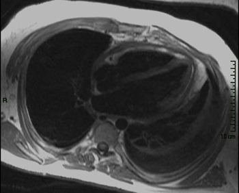
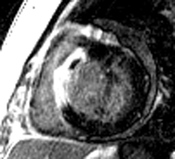
3) This horizontal long axis SSFP demonstrates which of the following entities?


Apical septum wall motion abnormality
Inferolateral wall motion abnormality
Global myocardial systolic dysfunction
Diastolic dysfunction


4) What are the primary physiologic consequences of the entity in the image shown below?


Decreased arterial pressures
Poor systolic function
Restricted diastolic filling and increase in venous pressures
Valvular regurgitation
Decreased venous pressures


5) The cine below shows findings which are characteristic of which of the following?


Dilated cardiomyopathy
Myocarditits
Arrhythmogenic right ventricular cardiomyopathy
Hypertrophic cardiomyopathy
Infiltrative cardiomyopathy


6) The gradient echo cine below shows what congenital abnormality?


Atrial septal defect
Ventricular septal defect
Tetralogy of Fallot
Transposition of the great vessels
Total anomalous pulmonary venous return


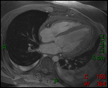
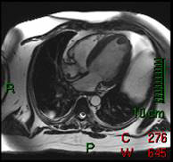
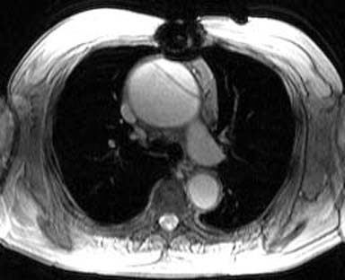
7) This spin echo image shows what aortic abnormality?


Aortic dissection
Aortic intramural hematoma
Aortic aneurysm
Aortic coarctation
Aortic coarctation


8) Which of the following is shown in the IR image below acquired 10 minutes following infusion of gadolinium contrast agent?


Transmural infarct
Subendocardial infarct
Myocarditis
Ventricular aneurysm
No abnormality


9) The cine image below uses a technique specialized to determine what entity?


Ejection fraction
Myocardial mass
Myocardial strain
Myocardial perfusion
Myocardial stress


10) Which congenital cardiac anomaly is shown on the gradient echo cine below?


Ebstein’s anomaly
Atrial septal defect
Tetralogy of Fallot
Ventricular septal defect
Pulmonic atresia


11) Below are pre- and post-gadolinium SSFP images. What abnormality do they show?


Microvascular obstruction
Myxoma
Cardiac metastasis
Thrombus
None, normal papillary muscle


12) Which is seen on the gradient echo cine below?


Pericardial effusion
Pericardial effusion causing cardiac tamponade
Arhythmogenic right ventricular cardiomyopathy
Pericardial cyst
Constrictive pericarditis


13) What abnormality is seen on this inversion recovery image?


Delayed hyperenhancement of infarction
Delayed hyperenhancement of myocarditis
Delayed hyperenhancement of hypertrophic cardiomyopathy
Dilated Cardiomyopathy
No abnormality


14) What abnormality is seen on the gradient echo vertical long axis cine seen below?


Myxoma
Intracardiac thrombus
Angiosarcoma
Lipoma
SVC thrombus


15) Below are perfusion images of basal, mid, and apical slices along with a delayed hyperenhancement image (bottom) aligned with the mid slice. The resting images are displayed on the top left with the Adenosine images displayed in the top right. What abnormality is seen in these images?


Artifact
Infarction of inferior wall
Infarction of anterior wall
Ischemia of inferior wall
Ischemia of anterior wall


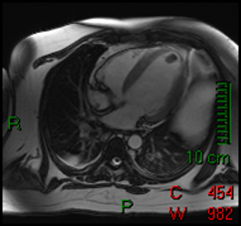
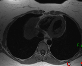
16) What aortic abnormality is seen on the T1 weighted axial image below?


Myocardial rupture
Descending aortic aneurysm
Aortic intramural hematoma
Stanford type A aortic dissection
Stanford type B aortic dissection


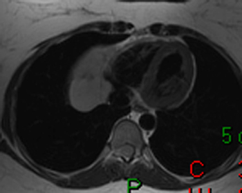
17) What abnormality is seen on the T1-weighted (left) and T2-weighted (right) images below?


Pericardial cyst
Myxoma
Pericardial mesothelioma
Constrictive pericarditis
Lipoma


18) This image is most consistent with what diagnosis?


Transmural infarction
Subendocardial infarction
Amyloidosis
Constrictive pericarditis
Ventricular septal defect


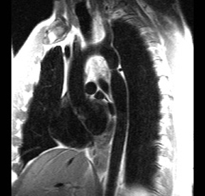
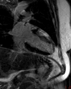
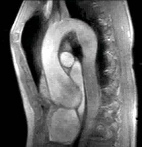
19) What abnormality is seen on the sagital view below?


Type A aortic dissection
Type B aortic dissection
Aneurysm of the aortic root
Aortic intramural hematoma
Aortic coarctation


20) What abnormality is seen in the DHE image below?


Sarcoidosis
Subendocardial infarction
Microvascular obstruction
Myocarditis
None, no DHE seen


Your score is out of 20.

Answers

© Copyright Rector and Visitors of the University of Virginia 2021