Cardiac MRI > Technique > MRI Physics
MRI Physics
![]()
Magnetic resonance (MR) imaging relies on positively charged hydrogen protons in the body, most of which are located in water molecules. When a patient is placed in a magnetic field, these protons begin to spin at a frequency that is proportional to the strength of the magnetic field, called the Larmor frequency. Each proton is a spinning positive charge, and this moving charge generates a small magnetic field. The small magnetic fields of the protons align themselves with the large magnetic field generated by the superconducting magnet.
A relatively small spatially varying magnetic field (gradient) is applied in addition to the large spatially constant magnetic field causing protons at different locations to rotate with slightly different frequencies. A radiofrequency (RF) energy pulse is then delivered by the RF coil to the protons. This RF pulse has the same frequency as the protons spinning in the desired imaging location. The RF pulse adds energy to only these protons by resonance; protons spinning at frequencies different from the RF pulse do not capture this energy. The energy from the RF pulse pushes the magnetization direction of the selected protons away from the direction of the large magnet field generated by the superconducting magnet.
When the radiofrequency pulse is stopped, the selected protons relax back to their original alignment with the large magnetic field. While relaxing they release a signal in the form of RF energy. This energy can be captured by an RF receiver coil and processed to yield localized information about the protons in the patient�s tissues. This process of disturbing selected protons and then collecting the energy released as the protons relax is the basis of MR imaging.
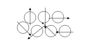
The circles represent hydrogen atoms and the arrows represent the directions of the magnetization vectors before the protons are exposed to the large uniform magnetic field of the MRI scanner.
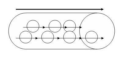
When placed in a MRI scanner, the magnetization vectors of the hydrogen atoms align themselves with the magnetic field. The large arrow on top represents the direction of the large magnetic field.
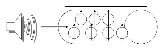
A radiofrequency pulse is delivered which causes the magnetization vectors of the protons to change direction.
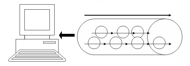
As the protons relax, they emit a radiofrequency pulse which is captured and an image is produced.
![]()
![]()