Cardiac MRI > Technique > Imaging of Flowing Blood
Imaging of Flowing Blood
![]()
Velocity encoded gradient echo imaging (VENC), also known as phase contrast imaging, is an MR technique for quantifying flowing blood. By measuring the phase shift that occurs as protons in the blood move through a magnetic field, the velocity and direction of the blood can be obtained. To obtain accurate measurements from a VENC image, a plane must be selected that is perpendicular to the path of the flowing blood. The technique creates a velocity encoded image for multiple phases of the heart cycle, thus creating a cine. In the cine produced, stationary tissue appears gray with tissue moving through the plane appearing as shades of either white or black, depending on the direction. The more white or black the tissue is, the faster it is moving. Quantification of blood flow is performed by software that requires the user to outline the vessel of interest for each phase. The software produces time-velocity and time-flow curves.
This technique is valuable for evaluating stenoses. A pressure gradient across a lesion can be calculated from blood velocities. Blood flow is quantified from the individual blood velocities at each point over the cross section of the blood vessel. This technique can be employed to determine the relative flows in the systemic and pulmonary systems when evaluating a cardiac shunt, determining the pressure gradient across a stenotic valve, or the regurgitant flow through a valve.
The figure on the left is a gradient echo cine of a patient with pulmonary valve regurgitation. A perpendicular line is drawn through the proximal pulmonary artery to designate the plane for the VENC cine on the right. During systole, there is white signal due to blood flowing towards the pulmonary arteries. However, as the cardiac cycle progresses, the signal becomes black in diastole due to the regurgitant blood flowing back into the right ventricle.
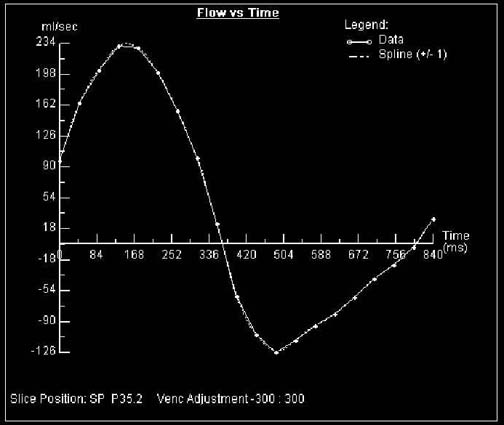
The figure shows a graphic representation of flow in the pulmonary artery. It is positive early in the cardiac cycle, but becomes negative during diastole due to regurgitant flow.
![]()
![]()