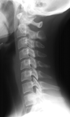
Imaging of the Cervical Spine > Interpretation > Bony Landmarks (cont.)
Bony Landmarks
![]()
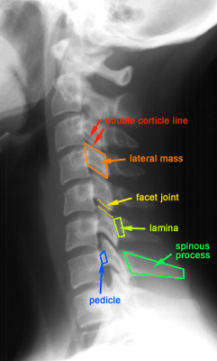
Pedicles project posteriorly to support the articular pillars, forming the superior and inferior margins of the intervertebral foramen. The left and right pedicels should superimpose on true lateral views. If fracture is suspected, get oblique views or CT.
Facets: the articular pillars are osseous masses connected to the posterolateral aspect of vertebral bodies via the pedicles. The facet joints are formed between each lateral mass. On the lateral view, the lateral masses appear as rhomboid-shaped structures projecting downward and posterior. "Double cortical lines" results from slight obliquity from lateral projection. The distance of the joint space should be roughly equal at all levels.
Lamina: the posterior elements are seen poorly on the lateral film. They are best demostrated by CT.
Spinous process: generally get progressively larger in the lower vertebral bodies. The C7 cervical spine is usually the largest.
![]()
![]()