Chest Radiology > Pathology > Emphysema & COPD
Emphysema
![]()
Emphysema is loss of elastic recoil of the lung with destruction of pulmonary capillary bed and alveolar septa. It is caused most often by cigarette smoking and less commonly by alpha-1 antitrypsin deficiency. Functional hallmarks are decreased airflow (decreased FEV1) and diffusing capacity (decreased DLCO2).
Emphysema is commonly seen on CXR as diffuse hyperinflation with flattening of diaphragms, increased retrosternal space, bullae (lucent, air-containing spaces that have no vessels that are not perfused) and enlargement of PA/RV (secondary to chronic hypoxia) an entity also known as cor pulmonale. Hyperinflation and bullae are the best radiographic predictors of emphysema. However, the radiographic findings correlate poorly with the patient's pulmonary function tests. CT and HRCT (high resolution CT) has emerged as a technique to evaluate different types, panlobular, intralobular, paraseptal and for guidance prior to volume reduction surgery.
Occasionally the trachea is very narrow in the mediolateral plane in emphysema. "Saber sheath" tracheal deformity is when the coronal diameter is less than 2/3 that of the sagittal.
In smokers with known emphysema the upper lung zones are commonly more involved than the lower lobes. This situation is reversed in patients with alpha-1 anti-trypsin deficiency, where the lower lobes are affected.
Chronic bronchitis commonly occurs in patients with emphysema and is associated with bronchial wall thickening.

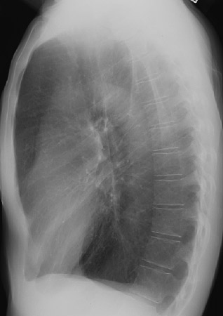
Note bilateral flattening of the diaphragms and significant hyperinflation as demonstrated by visualization of 11 posterior ribs.
![]()
![]()