Chest Radiology > Pathology > Atelectasis > General
Atelectasis
![]()
Atelectasis is collapse or incomplete expansion of the lung or part of the lung. This is one of the most common findings on a chest x-ray. It is most often caused by an endobronchial lesion, such as mucus plug or tumor. It can also be caused by extrinsic compression centrally by a mass such as lymph nodes or peripheral compression by pleural effusion. An unusual type of atelectasis is cicatricial and is secondary to scarring, TB, or status post radiation.
Atelectasis is almost always associated with a linear increased density on chest x-ray. The apex tends to be at the hilum. The density is associated with volume loss. Some indirect signs of volume loss include vascular crowding or fissural, tracheal, or mediastinal shift, towards the collapse. There may be compensatory hyperinflation of adjacent lobes, or hilar elevation (upper lobe collapse) or depression (lower lobe collapse). Segmental and subsegmental collapse may show linear, curvilinear, wedge shaped opacities. This is most often associated with post-op patients and those with massive hepatosplenomegaly or ascites .

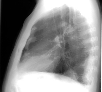
Note the loss of the right heart border silhouette due to partial atelectasis of the RML. Atelectasis is usually, but not always, a benign finding as in this example which was caused by an endobronchial mass in the RML.


This is a PA and lateral film showing round atelectasis, where the lung becomes attached to the chest wall by an area of previous inflammation. The lung then rolls up, causing this opacity.
