Chest Radiology > Pathology > Atelectasis > Left Lung
Left Lung Atelectasis
![]()
Left Upper Lobe
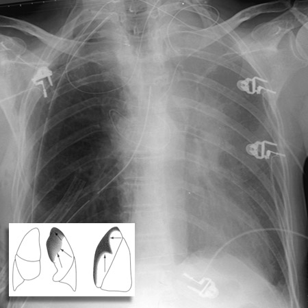
The left lung lacks a middle lobe and therefore a minor fissure, so left upper lobe atelectasis presents a different picture from that of the right upper lobe collapse. The result is predominantly anterior shift of the upper lobe in left upper lobe collapse, with loss of the left upper cardiac border. The expanded lower lobe will migrate to a location both superior and posterior to the upper lobe in order to occupy the vacated space. As the lower lobe expands, the lower lobe artery shifts superiorly. The left mainstem bronchus also rotates to a nearly horizontal position.

This patient suffered from left upper lobe atelectasis following right upper lobectomy.


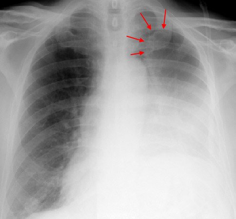
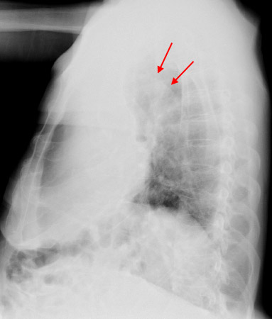
PA and Lateral of a patient with Left Upper Lobe Collapse (arrows). This characteristic finding on CXR is known as the Luftsichel Sign and may represent collapse due to obstruction from a bronchogenic carcinoma. The lucency between the mediastinum and the collapsed LUL is caused by hyperexpansion of the superior segment of the LLL.
Left Lower Lobe
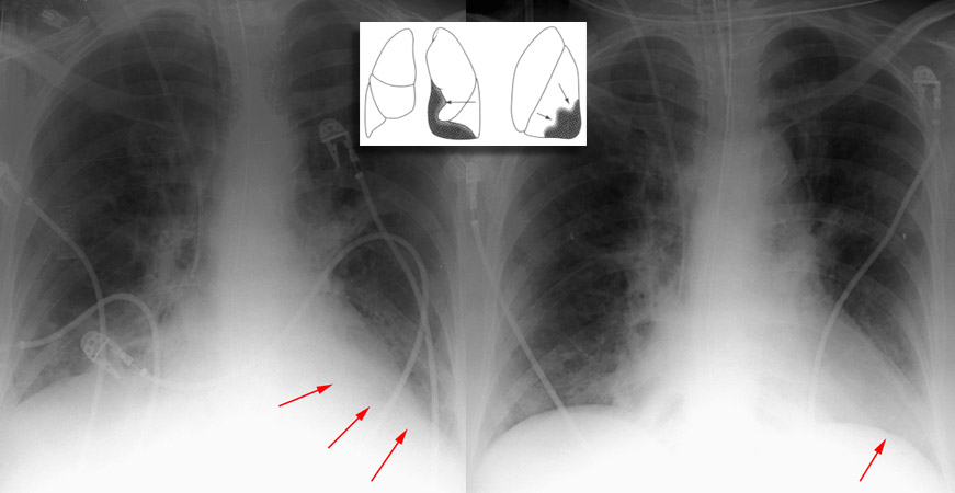
Atelectasis of either the right or left lower lobe presents a similar appearance. Silhouetting of the corresponding hemidiaphragm, crowding of vessels, and air bronchograms are sometimes seen, and silhouetting of descending aorta is seen on the left. It is important to remember that these findings are all nonspecific, often occuring in cases of consolidation, as well. A substantially collapsed lower lobe will usually show as a triangular opacity situated posteromedially against the mediastinum.

These radiographs demonstrate left lower lobe atelectasis followed by partial resolution, respectively.

Another PA exam of LLL atelectasis (arrows). Note the elevation of the left hemidiaphragm.
© Copyright Rector and Visitors of the University of Virginia 2013
