Chest Radiology > Pathology > Solitary Pulmonary Nodule
Solitary Pulmonary Nodule
![]()
A solitary pulmonary nodule is not an uncommon finding on a chest x-ray. A solitary pulmonary nodule can result from a wide range of causes. Most nodules are benign but some can be malignant.
Nodules are diagnosed as benign if they:- Show little or no growth for 2 years
- Calcification
- Central, laminated or diffuse pattern indicates a granuloma
- Eccentric calcification can be seen in a carcinoma or in a cancer that has engulfed a granuloma
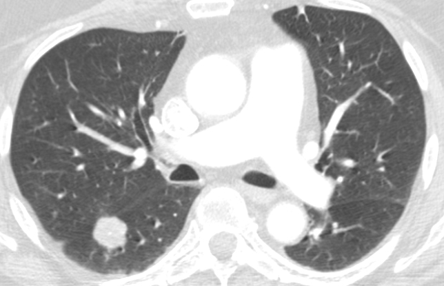
The CT image above shows a right lower lobe centrally calcified nodule, consistent with a benign granuloma.
A differential of possible etiologies is as follows:
- Granuloma - usually caused by fungal infections like histoplasmosis or tuberculosis
- Lung Carcinoma
- Solitary metastasis - usually from colon, breast, kidney, ovary, or testis
- Round pneumonia
- Abscess
- Round atelectasis
- Hamartoma - popcorn calcification is sometimes seen
- Sequestration
- Arteriovenous malformation
Other things can cause an apparent nodule but are actually outside the lung including:
- Fluid in an interlobar fissure
- Pleural plaques - small, often calcified, plate-like surfaces on the pleura often caused by asbestos fibers that invade the pleura from the lungs
- Skin lesions - nipple shadow, mole, lipoma, etc.
Low Risk Patient
| ≤ 6mm | No follow-up needed |
| 6-8mm | CT at 6-12 mo |
| >8mm | CT at 3 mo, PET CT or biopsy |
High Risk Patient (eg. smoking history or history of malignancy)
| ≤ 6mm | Optional CT at 12 mo |
| 6-8mm | CT at 6-12mo; consider- follow-up at 18-24 mo |
| >8mm | Consider CT at 3 mo, PET CT or biopsy |
![]()
![]()
© Copyright Rector and Visitors of the University of Virginia 2013