Chest Radiology > Pathology > Pleural Mass
Pleural and Extra-pleural Masses
![]()
The differential for pleural mass includes; metastases (especially adenocarcinoma and malignant thymoma), loculated pleural effusions (pseudotumor), malignant mesothelioma, pleural plaques from asbestosis (bilateral densities), and lymphoma. The differential for extrapleural mass includes rib tumor, rib infection (including chest wall fungal infection), neurofibroma or schwannoma (may erode a rib, but does not destroy it), and lipoma. One must first determine whether a mass arises from inside the lung or outside, an oblique margin with lung tissue indicates that the process is pleural or extrapleural. Distinguishing between a pleural and extrapleural mass can be challenging. If the center of the lesion is inside the chest wall, a pleural process is likely. Rib destruction indicates extrapleural involvement and possibly the origin of the mass.


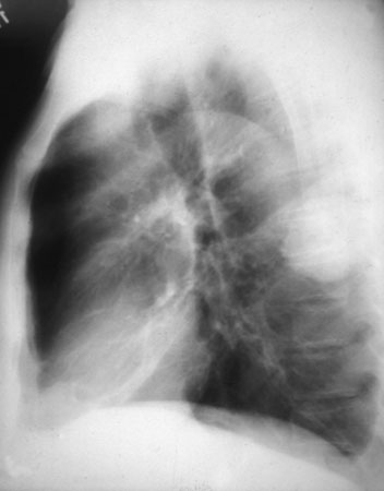
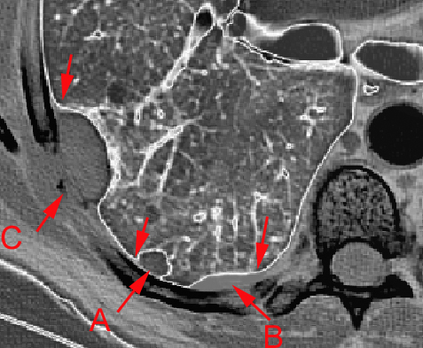
Lateral exam of an intraparenchymal mass. Note acute margins like "A" in the diagram on the right. Both "B" and "C" have obtuse margins. "B" demonstrates a pleural mass while "C" is an extrapleural chest wall mass.
© Copyright Rector and Visitors of the University of Virginia 2013
