Chest Radiology > Pathology > Tuberculosis
Tuberculosis
![]()
Primary tuberculosis (TB) is the initial infection with Mycobacterium tuberculosis. Post-primary TB is reactivation of a primary focus, or continuation of the initial infection. Radiographically, TB is represented by consolidation, adenopathy, and pleural effusion. A Ghon focus is an area of consolidation that most commonly occurs in the mid and lower lung zones. A Ghon complex is the addition of hilar adenopathy to a Ghon focus.
Radiographic features of post-primary TB are; focal patchy airspace disease "cotton wool" shadows, cavitation, fibrosis, nodal calcification, and flecks of caseous material. These occur most commonly in the posterior segments of the upper lobes, and superior segments of the lower lobes.
Endobronchial TB involves the wall of a major bronchus. Complications of endobronchial TB are cicatrical stenosis and obstruction.
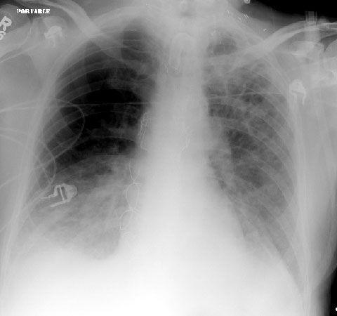
This is a PA exam of a patient who has had tuberculosis for
years.
This shows fibrosis, cavitation, and calcification, particularly in the left
upper lobe.
![]()
![]()
© Copyright Rector and Visitors of the University of Virginia 2013