Chest Radiology > Pathology > Pulmonary Edema > Pulmonary Edema
Pulmonary
Edema
![]()
There are two basic types of pulmonary edema. One is cardogenic edema caused by increased hydrostatic pulmonary capillary pressure. The other is termed noncardogenic pulmonary edema, and is caused by either altered capillary membrane permeability or decreased plasma oncotic pressure.
A helpful mnemonic for noncardiogenic pulmonary edema is NOT CARDIAC (near-drowning, oxygen therapy, transfusion or trauma, CNS disorder, ARDS, aspiration, or altitude sickness, renal disorder or resuscitation, drugs, inhaled toxins, allergic alveolitis, contrast or contusion.
On a CXR, cardiogenic pulmonary edema can show; cephalization of the pulmonary vessels, Kerley B lines or septal lines, peribronchial cuffing, "bat wing" pattern, patchy shadowing with air bronchograms, and increased cardiac size. Unilateral, miliary and lobar or lower zone edema are considered atypical patterns of cardiac pulmonary edema. A unilateral pattern may be caused by lying preferentially on one side. Unusual patterns of edema may be found in patients with COPD who have predominant upper lobe emphysema.
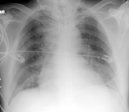
PA exam of a patient with pulmonary edema showing cephalization of pulmonary veins and indistinctness of the vascular margins. The heart is enlarged. |

Would you favor pneumonia or
CHF in this patient? Why? What pattern is shown?
(Click image for answer)
![]()
![]()
Above are two exams from the same patient. The left radiograph clearly shows diffuse pulmonary edema with loss of both hemidiaphragms and silouhetting of the heart. The exam on the right was taken two days later after partial resolution of the edema.
![]()
![]()
© Copyright Rector and Visitors of the University of Virginia 2013