Chest Radiology > Interpretation > Signs > Air Bronchogram
Air
Bronchogram
![]()
An air bronchogram is a tubular outline of an airway made visible by filling of the surrounding alveoli by fluid or inflammatory exudates. Six causes of air bronchograms are; lung consolidation, pulmonary edema, nonobstructive pulmonary atelectasis, severe interstitial disease, neoplasm, and normal expiration.

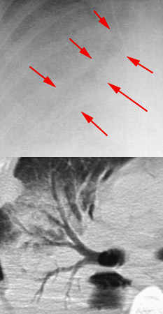
This patient has bilateral lower lobe pulmonary edema. The alveoli are filled with fluid making the bronchi visible as an air bronchogram. The upper right is a closeup of the right side of the image with arrows outlining a prominent air bronchogram. The lower right is a CT scan demonstrating an air bronchogram clearly.
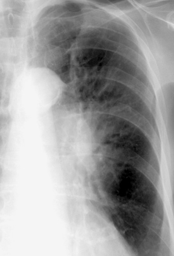
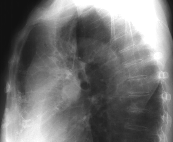
Here is another example of air bronchograms in both a PA and Lateral exam. Can you spot them?
![]()
![]()
© Copyright Rector and Visitors of the University of Virginia 2013