Chest Radiology > Pathology > Hilar Adenopathy
Hilar Adenopathy
![]()
Enlargement of the lymph nodes within the lung hilum can be an important finding for underlying pathology.
A differential of possible etiologies can be broken up into three different categories:- Inflammation (sarcoidosis, silicosis)
- Neoplasm (lymphoma, metastases, bronchogenic carcinoma)
- Infection (tuberculosis, histoplasmosis, infectious mononucleosis)
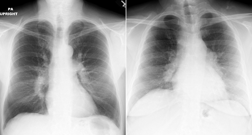
An important consideration to keep in mind is that since the pulmonary arteries also course through the same area, enlargement of these vessels may be confused with hilar adenopathy. Typically, lymphadenopathy has a more lumpy-bumpy appearance, while an enlarged pulmonary artery appears smooth.
One of the following images displays hilar adenopathy and the other shows pulmonary artery enlargement. Can you determine which one is which?

The above left image shows bilateral pulmonary artery enlargement. Note the smooth contours of the arteries. The above right image shows the lumpy-bumpy opacities characteristic of hilar adenopathy.
© Copyright Rector and Visitors of the University of Virginia 2013
