Chest Radiology > Pathology > Pericardial Effusion
Pericardial
Effusion
![]()
Pericardial effusion causes an enlarged heart shadow that is often globular shaped (transverse diameter is disproportionately increased). A "fat pad" sign, a soft tissue stripe wider than 2mm between the epicardial fat and the anterior mediastinal fat can be seen anterior to the heart on a lateral view. Serial films can be helpful in the diagnosis especially if rapid changes in the size of the heart shadow are observed. Approximately 400-500 ml of fluid must be in the pericardium to lead to a detectable change in the size of the heart shadow on PA CXR. Pericardial effusion can be definitively diagnosed with either echocardiography or CT. It can be critical to diagnose pericardial effusion because if it is acute it may lead to cardiac tamponade, and poor cardiac filling. In the postoperative patient it could be a sign of bleeding, necessitating a return to the OR.
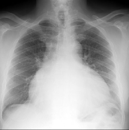
PA of a patient with a pericardial effusion.
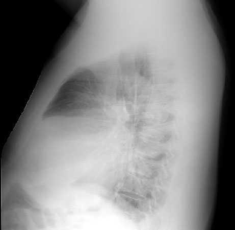
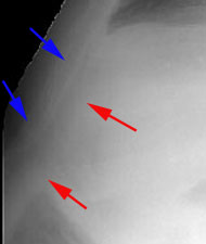
A lateral exam and closeup of a pericardial effusion showing the
anterior mediastinal fat (blue arrows)
and epicardial fat (red arrows) separated
by a soft tissue stripe reflecting the pericardial effusion seen edge-on.
![]()
![]()
© Copyright Rector and Visitors of the University of Virginia 2013