Chest Radiology > Interpretation > Signs > Silouhette Sign
Signs
![]()
Silhouette sign
One of the most useful signs in chest radiology is the silhouette sign. This was
described by Dr. Ben Felson. The silhouette sign is in essence elimination of
the silhouette or loss of lung/soft tissue
interface caused by a mass or fluid in the normally air filled lung. In other
words, if an intrathoracic opacity is in anatomic contact with, for example,
the heart border, then the opacity will obscure that border. The sign is
commonly applied to the heart, aorta, chest wall, and diaphragm. The location of
this abnormality can help to determine the location anatomically.
Take a moment to review the makeup of the mediastinal
margins and the lobes of the lungs that interface with the mediastinum.
Use the back button on your browser to return here.
For the heart, the silhouette sign can be caused by an opacity in the RML, lingula, anterior
segment of the upper lobe, lower aspect of the oblique fissure, anterior mediastinum, and anterior portion of the pleural cavity. This contrasts with an
opacity in the posterior pleural cavity, posterior mediastinum, of lower lobes
which cause an overlap and not an obliteration of the heart border. Therefore
both the presence and absence of this sign is useful in the localization of
pathology.
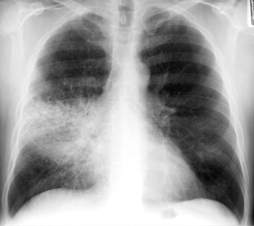
The right heart border is silhouetted out.
This is caused
by a pneumonia, can you determine which lobe the pneumonia affects?
(click
image for answer)

