Chest Radiology > Pathology > Mediastinal Mass > Posterior Mediastinum
Posterior Mediastinal
Mass
![]()
The differential for a posterior mediastinal mass includes; neoplasm, lymphadenopathy, aortic aneurysm, adjacent pleural or lung mass, neurenteric cyst or lateral meningocele, and extramedullary hematopoiesis.

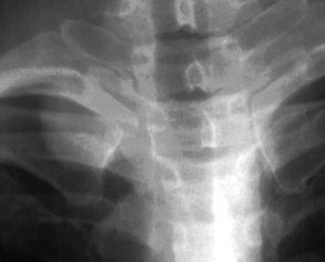
Note that this mass is detected by a pleural margin search as you move your eye along the superomedial part of the right lung. The interface is interrupted. Think about the anatomy of the lung in this area. The anterior mediastinum ends at the level of the clavicles. Any abnormality in the apex of the thorax must be posterior in the chest.

The mass projects above the clavicles, therefore it is not an anterior structure.


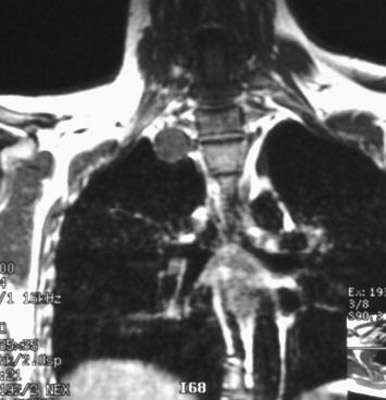
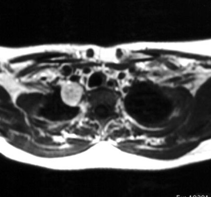
This MRI shows the mass is extrapleural and associated with the spinal nerves. It is a schwannoma, a benign tumor of the nerve sheath.
