Emergency Ultrasound > Epigastric Pain > AAA
Epigastric Pain - AAA
![]()
Clinical
Approximately 90-95% of abdominal aortic aneurysms (AAA) are confined to the infrarenal aorta. AAA are usually not repaired until they exceed 4-5 cm in maximum diameter. The risk of rupture within 5 years is 25% at 5 cm diameter. AAA smaller than 5 cm have a 3% risk of rupture over 10 years. US is used to monitor the rate of enlargement of AAA. The average increase is 2 mm/yr diameter.
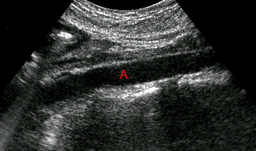
Longitudinal image through the normal abdominal aorta (A) with a diameter of 2 cm.
Exam
Begin with the patient in the supine position. Obtain longitudinal and transverse images of entire abdominal aorta and a transverse view of bifurcation to show the iliac arteries. Get a longitudinal image of each iliac artery. Image the superior mesenteric artery (SMA) and celiac artery. Image renal arteries if origins are seen.
AAA measurements: AP measurement in longitudinal and transverse views. Measure transverse diameter. Measurements are outer wall to outer wall. If AAA is found, obtain coronal views of right and left kidneys for renal length.
Sonographic Findings:
1) Abdominal aorta > 3 cm, measured from inner wall to inner wall.
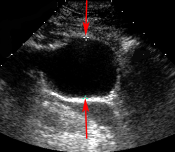
Transverse image demonstrating focal enlargement of the aorta (red arrows) with a diameter of 4 cm.
2) Hypoechoic mural thrombus within AAA.
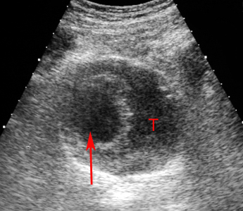
Enlarged view of the right common iliac artery shows the large amount of intraluminal thrombus (T) commonly found in aneurysms of the aorta and iliac arteries. The patent lumen is indicated by the red arrow.
3) Rupture of AAA is suggested by fluid or hematoma around the aorta.
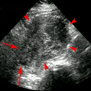
Transverse image demonstrates rupture of an aortic aneurysm (red arrows) while the red arrowheads indicate the intact aortic wall.
![]()
![]()