Emergency Ultrasound > Technique > Scanner
Technique - Scanner
![]()
The electrical signals are sent to the scanner from the transducer and analyzed to produce an image. The image is a result of the strength of the echo (brightness=sound intensity) and the time at which the echo is received. The ultrasound image is displayed as tiny white pixels on a black background. The gray-scale image can portray structures from a spectrum of anechoic to hyperechoic. Anechoic or echolucent structures have complete absence of echoes and therefore appear black. Hyperechoic or echogenic structures have more echoes (whiter) than surrounding tissue.
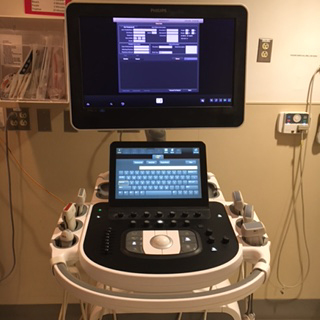
Image of a scanner.
A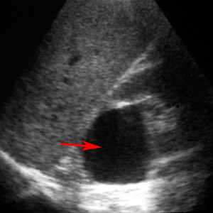 B
B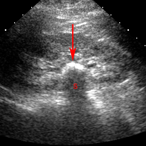
A. Anechoic structure (fluid filled cyst) indicated by red arrow. B. Hyperechoic structure (renal stone) indicated by red arrow produces an acoustic shadow (S).
Transverse images are displayed with the patient's right side on the left side of the the image like a CT scan. Longitudinal images are displayed with the cranial aspect of the patient's anatomy at the left side of the image.
A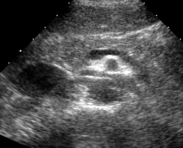 B
B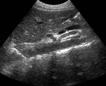
A. Transverse image of normal abdomen. B. Longitudinal image of normal abdomen.
![]()
![]()