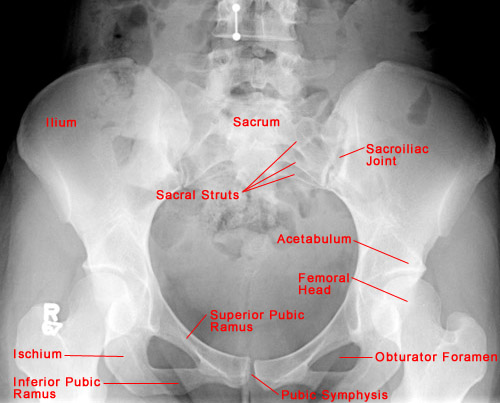
- The five bones that comprise the pelvis are the ilium, ischium, pubis, sacrum, and
coccyx.
- Most trauma to the pelvis and hips can be evaluated with an AP projection of the pelvis
and hips. Other injuries require special projections such as anterior and posterior obliques
views of the pelvis, frog-lateral view of the hip and groin-lateral view.
- CT of the pelvis is the technique of choice for evaluating complex fracture patterns,
degree of displacement and soft tissue injury.
- Symptoms from fractures of the hip, acetabulum and pelvis may be quite similar, thus, a full AP pelvis
radiograph including the hip must be obtained if any of the above fractures are expected.
- The femurs should be internally rotated when obtaining an AP pelvis film so that the femoral necks can be
appropriately assessed for fractures.
|