|
The following radiographs show the normal anatomy of the ankle.
|

|
Lateral View of Normal Ankle
|
|
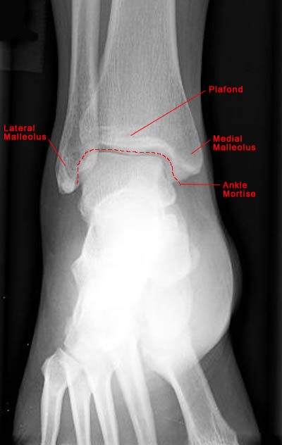
AP View of Normal Ankle
|
|
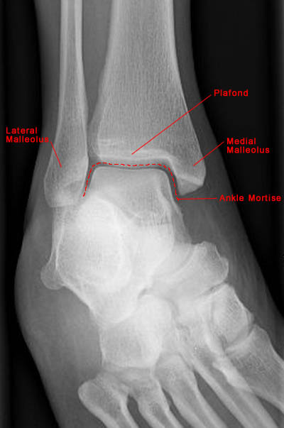
Mortise View of Normal Ankle
|
- The ankle joint includes articulations between three structures: tibia, fibula and talus.
- The lateral malleolus is longer than the medial malleolus, a characteristic important
for joint stability.
- Movement at the ankle joint is limited to dorsiflexion and plantarflexion. However, ankle
movement is typically combined with movement of joints in the foot resulting in many ranges of motion.
- Standard radiographic examination of the ankle includes AP, lateral and mortise views of the joint.
- Mortise view requires oblique examination at 20 to 30 degrees internal rotation and results in alignment
of medial and lateral malleoli in the same plane.
|
|
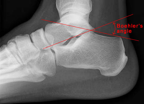
Lateral view of ankle showing Boehler's angle
|
- Boehler's angle is defined as the angle formed by two lines: one drawn tangent to the superior aspect of the
calcaneus and the second drawn tangent to the inferior aspect of the calcaneus. The angle normally ranges from 20 to
40 degrees.
|