Calcaneal Fracture
- Also known as a "lover's fracture," fractures of the calcaneus are usually the result of a
fall from height.
- A Boehler's angle less than 20 degrees is a characteristic sign of calcaneal fracture.
However, it is important to realize that compression fracture is not excluded by a normal
Boehler's angle.
- In 10% of cases, calcaneal fractures occur bilaterally.
- When evaluating a calcaneal fracture, it is important to determine whether the fracture line
involves the subtalar joint. CT is usually essential in performing this evaluation.
- Calcaneal fractures can often be associated with spinal compression fractures and fractures of
the femoral necks and tibial plateaus. For this reason, films of the thoracic and lumbar spine,
tibial plateaus and femoral necks may be required for the patient with a calcaneal fracture in order
to exclude other fractures.
|
|
|
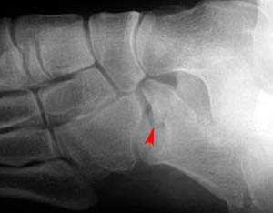
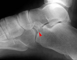
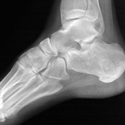
Lateral views of calcaneal fractures
|
|
Now look for the calcaneal fracture (and other abnormalities) in the image below.
Can you find them? Click on the image for the answer.
|
|
Lateral view
|
|
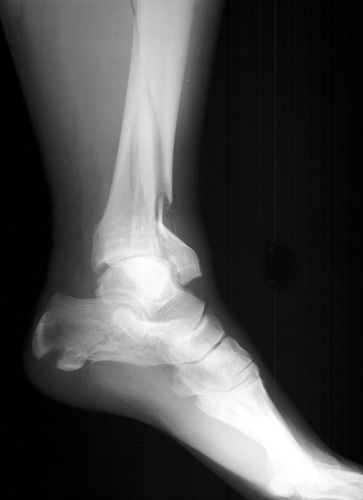
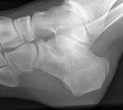
In addition to the anterior and posterior calcaneal fractures shown in the images above,
compression and stress fractures of the calcaneus are also common. Look at the two images
below showing compression fractures of the calcaneus.
|
|
|
Posterior tangential view
|
Lateral view
|
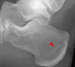
Stress fractures of the calcaneus are similar to stress fractures seen in other parts of the body.
- Fracture is not apparent immediately following the precipitating incident. Instead, the fracture
is usually seen between 10 and 14 days after its occurence.
- The fracture is seen as a sclerotic band on radiograph.
- When a stress fracture is suspected, but not visible on plain film, a bone scan may be used to confirm diagnosis.
- In cases of calcaneal stress fracture, the fracture line is usually vertical or parallel to the posterior contour
of the bone.
- Calcaneal stress fractures are most common in runners, but also occur in elderly indiviuals who
have weakened bones due to osteoporosis.
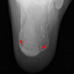
Look at the following radiographs showing stress fractures of the calcaneus.
|
|
|
Oblique view
|
Close-up of lateral view
|
;)