GI Radiology > Esophagus > Functional Abnormalities
Functional Abnormalities of the Esophagus
![]()
Achalasia |
|
Clinical Achalasia is a primary
esophageal motility disorder characterized by aperistalsis and lower esophageal
dysfunction. It is thought to be a neurogenic disorder. Clinically, patients
may complain of slowly progressive dysphagia for both solids and liquids.
Regurgitation of undigested food is also common and can lead to pulmonary
complications like coughing, aspiration or pneumonia. It occurs equally
in males and females during the middle decades (age 20 to 40) of life.
If these symptoms are present in an older patient and are accompanied
by chest pain, one should consider secondary achalasia due to malignancy.
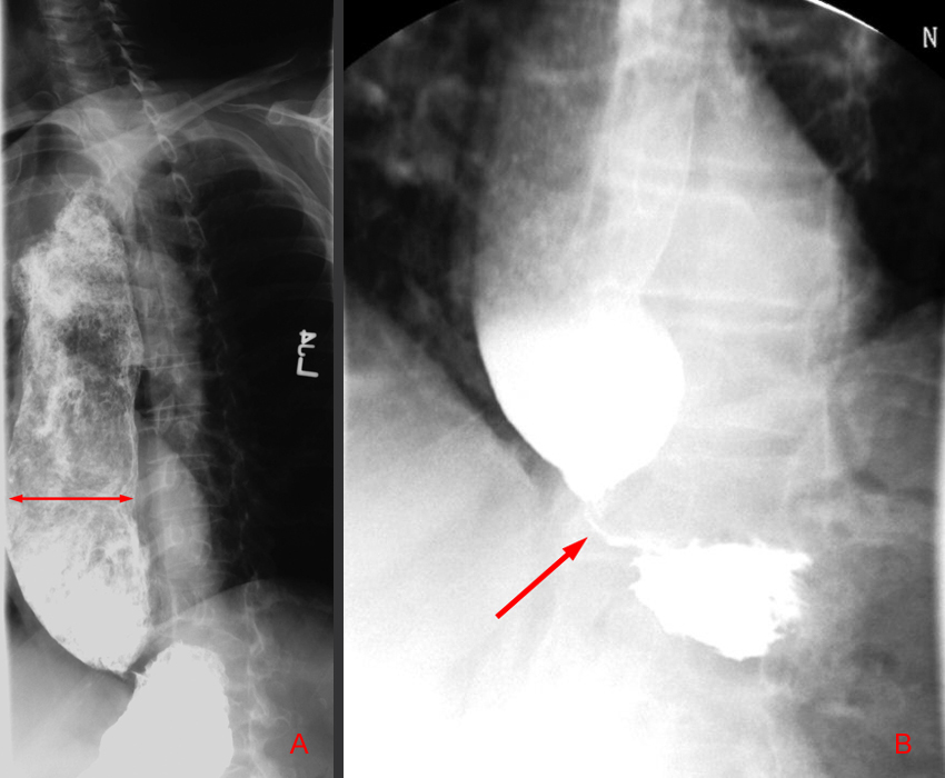
Radiological findings On plain films, achalasia can be recognized by massive esophageal dilatation with possible large amounts of retained food and fluid in the esophagus. The dilatation and tortuosity of the esophagus may present as a widened mediastinum with an air-fluid level. On contrast studies, the esophagus usually exhibits weak, nonpropulsive, and dysrhythmic peristaltic waves. The exam should be performed in the recumbent position because in the upright position, gravity can simulate the effects of peristalsis. Typically, the distal esophagus has a smooth, tapered appearance often likened to a "bird's beak" which reflects the LES dysfunction.
Image "A" depicts a lateral view of the esophagus showing a massively dilated esophagus with retention of contrast in the distal portions of the esophagus. Image "B" shows the "bird's beak" appearance of the dysfunctional lower esophageal sphincter.
Treatment Achalasia is often
treated by pneumatic dilatation of the LES or myotomy. Treatment is often
complicated by reflux esophagitis and subsequent peptic strictures.
|