- The hepatic veins lie between segments
whereas the portal vein lies within segments. Exception: the left portal
vein does not lie within segments. These vessels play an important
radiological role because they aid radiologists in approximately
localizing a particular lesion to the correct anatomical section of the
liver.
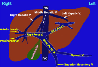
AP View of the Liver (3D)
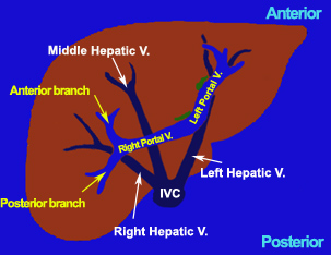
Axial View of the Liver (3D)
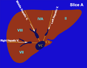
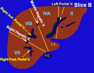
- Cephalad in the liver, above the level
of the main portal vein:
- The middle hepatic vein separates the
right lobe from the left lobe, or to be more specific, the right lobe,
superior anterior subsegment (VIII), from the left lobe, superior medial
subsegment (IVA).
- The right hepatic vein separates the
superior anterior (VIII) and superior posterior (VII) subsegments of the right lobe.
- The left hepatic vein separates the
superior medial (IVA) and superior lateral (II) subsegments of the left lobe.
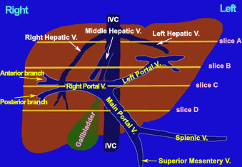
AP View of the Liver (3D)
Axial View of the Liver (2D)
- At the level just above the main portal vein:
- The left portal vein separates the
superior medial (IVA) subsegment from the superior lateral (II)
subsegment of the
left lobe.
AP View of the Liver (3D)
Axial View of the Liver (2D)
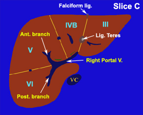
- Caudal in the liver, below the level of
the main portal vein:
- The line that bisects the angle
between the anterior and posterior branches of the right portal vein separates the
inferior anterior (V) and inferior posterior (VI) subsegments of the right lobe. First, you
need to identify the right portal vein. Then, the
anterior
branch will go into the inferior anterior
subsegment (V) while the
posterior branch will go towards the
inferior
posterior subsegment
(VI).
AP View of the Liver (3D)
Axial View of the Liver (2D)
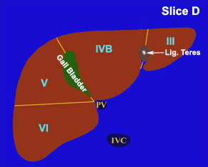
- The gallbladder separates the right
lobe, inferior anterior subsegment (V) from the left lobe, inferior medial
subsegment (IVB).
- The ligamentum teres divides the left
lobe into the inferior medial (IVB) and inferior lateral (III)
subsegments.
AP View of the Liver (3D)
Axial View of the Liver (2D) |