|
Focal fatty infiltration
- Pathogenesis:
- The low-density region on CT emulates a
low-attenuation metastasis.
- More common on either side of the falciform
ligament, in the vicinity of the gallblader fossa, and in the
posterior aspect of segment IV (left lobe).
- Associated with obesity, alcohol abuse,
steroids, and hyperalimentation.
- Radiographic findings:
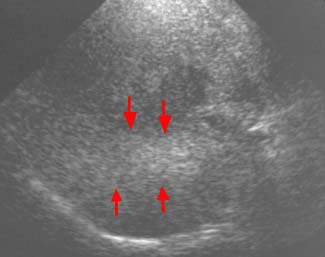
- U/S: bandlike geographic areas of
increased echogenicity in a lobar/segmental distribution (arrows).
- CT: no mass effect on adjacent vessels,
patchy areas of decreased attenuation.
- MRI:
can use "chemical shift" or "in-phase and
out-of-phase" gradient-echo imaging to detect focal fat
variation. This method is both highly sensitive and specific in
detecting fat and water in the same image voxel. On in-phase image,
fat and water combine their signals. On out-of-phase image, fat
and water signals cancel out, thus resulting in reduced relative
signal when both are present.
Fatty sparing
- Pathogenesis:
- A normal liver looks hyperdense to a
fatty infiltrated liver on noncontrast CT. In a fatty infiltrated liver,
there are areas that are spared from the infiltration process,
therefore, appearing "normal" or "hyperdense" to the surrounding fatty
liver. These hyperdense areas can create an appearance of pseudomasses.
- Characteristic locations for sparing
include the periportal regions, caudate lobe, and adjacent to the
gallbladder fossa.
- Radiographic findings:
- The lesion is hyperdense to the
surrounding liver parenchyma. The lesion is actually normal,
non-infiltrated liver tissue.
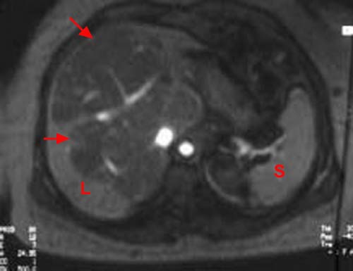
- MRI:
can use "chemical shift" or "in-phase and
out-of-phase" gradient-echo imaging to detect focal fat
variation. This method is both highly sensitive and specific in
detecting fat and water in the same image voxel. On in-phase image,
fat and water combine their signals. On out-of-phase image, fat
and water signals cancel out (arrows), resulting in reduced relative
signal when both are present. The image below is an example of chemial fat saturation.
Atypical regenerative nodules
-
Pathogenesis:
-
In chronic cirrhosis, some regenerative nodules
may resemble masses (i.e. hepatocellular carcinoma) on US
and MRI.
-
Radiologic
Findings:
-
US:
lesions may appear well-defined, homogeneous, and hypoechogenic.
-
Biphasic
CT: lesions are often not
visualized.
-
MRI:
they are often of increased signal on T1 weighting (due to accumulation
of fat or glycogen) and reduced signal on T2-weighted images (due to
accumulation of iron).
NOTE: malignant foci
can develop within
these nodules. On T2-weighted MRI, these foci appear as high
signals within the low signal nodules. Therefore, if there is
heterogeneity within nodules, malignancy should be suspected.
|