GI Radiology >
Liver >
Imaging Modalities >
Ultrasound
Modalities
of Liver Imaging

Ultrasound
|
- Is the first and the most commonly
obtained method of examination in patients with RUQ pains, abnormal LFTs, or
suspected liver masses.
- Is a noninvasive and excellent screening
tool.
- Used to evaluate the presence of bile
duct obstruction and gallstones as well as to distinguish a solid lesion
from a cystic one.
- Has low sensitivity and high false
negative rate for detection of liver metastases.
- 6 common causes of homogeneously
increased echogenicity of the liver are:
- Fatty infiltration
- Cirrhosis
- Hepatitis (sometimes)
- Amyloidosis
- Leukemic infiltration (often normal
to low echogenicity)
- Hemochromatosis
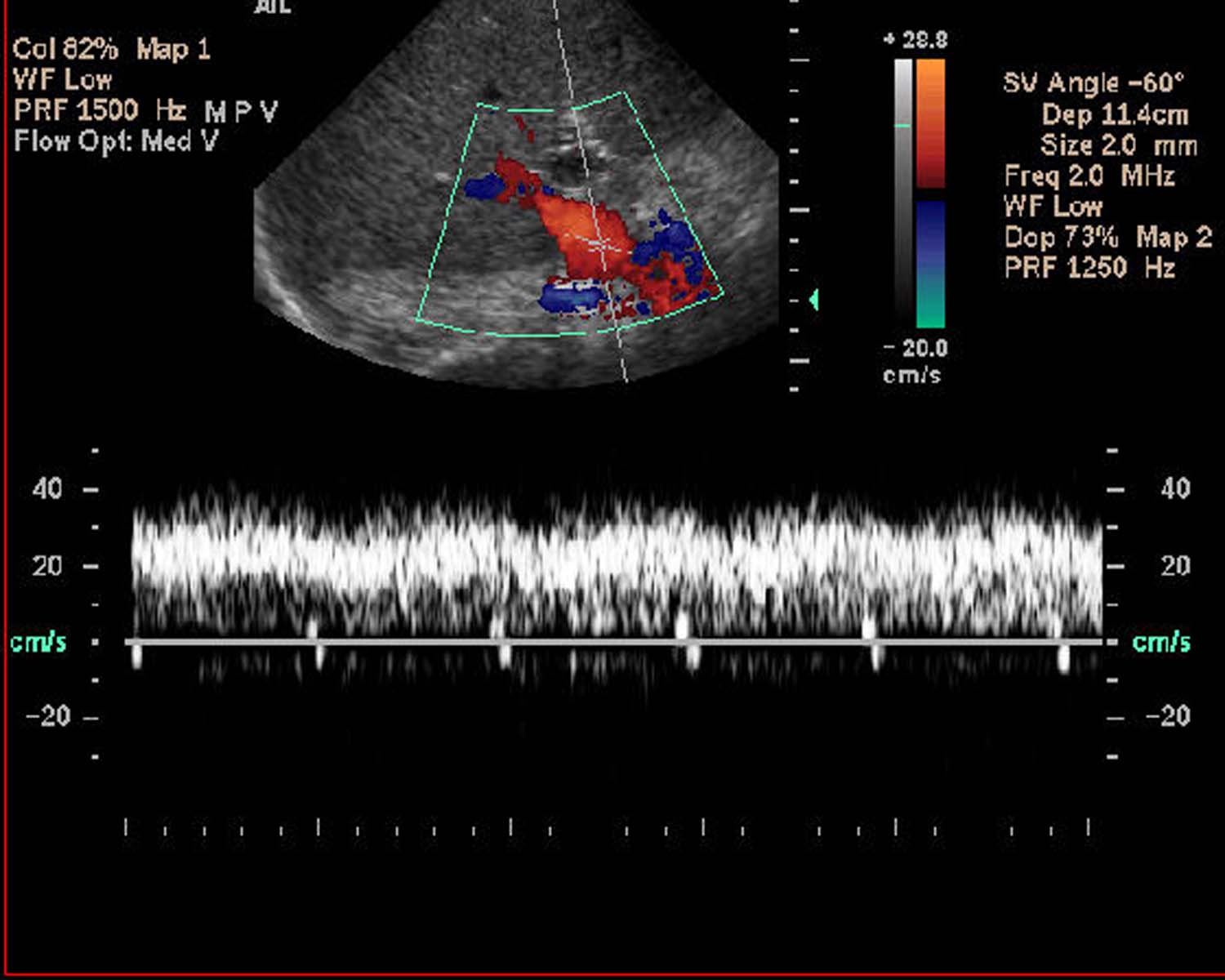
- Ultrasound Doppler imaging can be very
helpful in identifying vascular abnormalities, i.e. patency of hepatic
vessels, portal vein, and IVC as well as flow direction in these
vessels. Flow in the portal vein and hepatic arteries are hepatopedal
(toward the liver) while flow in hepatic veins and hepatic ducts are
hepatofugal (away from the liver).

- In color Doppler imaging, shades of red
typically refer to blood flowing toward the transducer, whereas shades
of blue refer to blood flowing away from the transducer.
- Commonly used terminology in U/S:
echogenic, anechoic, hyper/hypoechoic, acoustic shadow....
|
|


![]()
![]()