Pancreas divisum represents an anomaly of the fusion of the
two pancreatic buds and their respective ducts. It is the most common
pancreatic ductal system anomaly; it is seen in 4-6% of ERCP patients.
The duct of ventral bud does not fuse with the duct system
of the dorsal pancreatic bud; it joins the common bile duct at the level of the ampulla of Vater. The dorsal duct of
pancreas, which drains the anterior head, body
and tail, terminates more cephalad, at the minor duodenal papilla.
The narrow size of the os in the minor papilla may result in inefficient
drainage of pancreatic secretions. It is widely believed that
pancreas divisum predisposes to chronic pancreatitis.
|
|
Embryological
Development of the Pancreas |
|
Friedman AC: The pancreas.
In
Taveras & Ferrucci, Radiology,
Diagnosis-Imaging-Intervention.
Phila: Lippincott-Raven,
1992, p 2. |
C. Pancreas divisum
D. Normal fusion of the pancreatic buds and ducts |
|
|
Schematic
Representation of Normal and Divisum Ductal System |
|

Gay, SB. "Radiology Recall" Lippincott
Williams & Wilkins 2000, p394. |
|
ERCP is the best modality for making the diagnosis of pancreas
divisum. ERCP findings are described below. Endoscopic
interventions have yeilded encouraging results for the treatment of
pancreas divisum with acute recurrent pancreatitis. These
interventions include endoscopic sphincterotomy of
the minor ampulla with or without sphincterotomy of the major ampulla,
ductal balloon dilatation, and pancreatic duct stent placement.
|
|
ERCP Findings in
Pancreas Divisum |
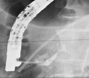 |
Cannulation of the major duodenal papilla and injection of contrast
material reveals only a short
segment of the main pancreatic duct that arborizes within the head of pancreas, and does not opacify
the main pancreatic duct in the pancreatic body and tail. A
stent inserted previously through the minor papilla indicates the
location of the dorsal pancreatic duct. |
 |
Cannulation of the minor duodenal papilla and opacification of the
dorsal pancreatic duct shows changes of chronic pancreatitis
(alternating dilation and strictures producing a beaded apprearence). |
|
Although the demonstration of ductal
anatomy by CT is very difficult, the use of thin slices and appropriate
algorithms can often show the pancreatic duct for most of its course.
CT diagnosis of pancreas divisum is the based on the failure to see the
union of the dorsal and ventral ducts. Additional CT signs of
pancreatitis may be present in the dorsal duct distribution.
MRCP can also be used to image the pancreatic duct. The study
can be repeated after secretin injection, and may demonstrate a
functional obstruction of the pancreatic duct via the minor os. |
|
|