|
The pancreas develops from dorsal and ventral diverticula that buds from the
primitive foregut. These two buds fuse after the rotation of the
duodenum adjoins the two structures. The ventral bud develops from the hepatic duct,
and forms the uncinate process and the posterior/inferior head of the
pancreas. The dorsal bud develops directly from the foregut, forming
the anterior head, body and tail of the pancreas.
The distal duct
system of the dorsal bud joins with that of the ventral bud to form the
main pancreatic duct (of Wirsung), which joins with the common bile duct
at the level of the ampulla of Vater.
The distal duct system of the dorsal bud may persist as the accessory
pancreatic duct (of Santorini), and empties into the duodenum at the
minor duodenal papilla. The "normal" short axis measurement of the
pancreas is 2.5 cm at the head, and 1.5 cm at the tail; there are normal
variations. In general, the pancreas decreases
with size with age.
|
|
|
Embryological
Development of the Pancreas |
|
Friedman AC: The pancreas. In Taveras & Ferrucci,
Radiology, Diagnosis-Imaging-Intervention. Phila: Lippincott-Raven,
1992, p 2. |
C. Pancreas divisum
D. Normal fusion of pancreatic buds and ducts |
|
|
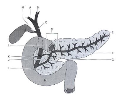
Pancreatic Anatomy |
|
Gay, SB. "Radiology Recall" Lippincott
Williams & Wilkins 2000, p392. |
A: Right hepatic duct
B: Left hepatic duct
C: Main hepatic duct
D: Main pancreatic duct (of Wirsung)
E: Pancreatic tail
F: Body of pancreas
G: Ligament of Treitz
H: Third part of duodenum
I: Major duodenal papilla/sphincter of Oddi
J: Head of pancreas
K: Accessory pancreatic duct (of Santorini)
L: common bile duct
M: Cystic duct |
|
Although the above described ductal anatomy is most common, there are
several congenital variations in the union of the two primitive
pancreatic ducts (the main ducts of the ventral and dorsal buds), as
illustrated below.
|
|
Common Variations in Ductal
Anatomy |
|
Friedman AC: The pancreas.
In Taveras
& Ferrucci, Radiology,
Diagnosis-Imaging-Intervention.
Phila: Lippincott-Raven, 1992, p 2. |
|
There are also several variations in the union of the bile and
pancreatic ducts, as illustrated below. It is important to be
aware of these variations in ductal anatomy,
which can be visualized using several imaging modalities.
|
|
Netter FH, Atlas of Human
Anatomy, CIBA-GEIGY, 1989, Plate 278 |
|
|
|