GI Radiology > Pancreas > Procedures

Procedures (Imaging Modalities)

   Imaging of the pancreas can be divided into two categories: indirect and direct imaging.  Indirect imaging involves non-invasive radiologic techniques including plain film, US, CT, MRI, and magnetic resonance cholangiopancreatography (MRCP).  Direct imaging involves invasive radiologic procedures, which include ERCP and operative pancreatography.


Indirect Imaging:
 

   Indirect imaging modalities of the pancreas include US, CT, MRI, and magnetic resonance cholangiopancreatography (MRCP).
 

Plain Films:

   The pancreas is not directly visualized on plain films of the abdomen.  Calcifications in the pancreatic region or biliary lithiasis can sometimes be demonstrated.  The presence of adynamic ileus of the doudenum and the proximal small bowel loops can be an important sign in acute disorders.

Ultrasonography (US):

   US is one method to evaluate the pancreas and peripancreatic structures.  The body of the pancreas is usually found anterior to the splenic vein.  In some patients however, body habitus and bowel gas may limit complete visualization. 
 
 
Computed Tomography (CT):

   CT pancreatography is used to evaluate the differences in the density, texture, and contour of the pancreas.  Secondary signs such as dilation of the bile or pancreatic duct, or changes in the peripancreatic fat density, are also used in the determination of a diagnosis.  Intravenous contrast enhancement is also used, especially in the analysis of pancreatic masses. 

Magnetic resonance imaging (MRI):


   MRI shows soft tissue with superb clarity, while avoiding some the potential risks of ionizing radiation.  Densities in an MR image are based on tissue water and lipid content
(while CT densities are based on varying absorption of X-rays by different tissue).


Magnetic resonance cholangiopancreatography (MRCP):

   MRCP is a non-invasive technique that delineates the pancreatic and biliary ductal systems, while providing projectional and cross sectional images of the ducts.  MRCP does not require administration of intravenous contrast material; it is based on T2-weighted images, which depict static fluid (including bile and pancreatic secretions), with a higher signal intensity.  MRCP also avoids the invasive complications of ERCP.  With the recent improvements in MRCP, it is superceding ERCP for many of its diagnostic indications.

   MRCP is inferior to ERCP in several respects however.  The spatial resolution of MRCP is lower than that of ERCP.  Furthermore, ascites or fluid collections in the upper abdomen can interfere with the visualization of the pancreatic and biliary ducts. 


Direct Imaging: 

   Direct imaging of the pancreas includes ERCP and operative pancreatography.


Endoscopic Retrograde Cholangiopancreatography (ERCP):

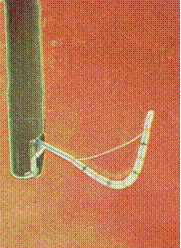
   ERCP is a combined endoscopic and radiographic procedure that images the biliary and pancreatic ducts.  ERCP is performed with a side-viewing duodenoscope which has an instrumentation channel that allows for the insertion of the cannulation catheter into the major or minor duodenal papilla.  A water-soluble contrast agent (60 % iodine) is injected into the ductal system using fluoroscopy for imaging.  It is important to obtain adequate ductal filling without over-distending the system.  (During interventional procedures, a guide-wire can be inserted through the cannulation catheter, for subsequent insertion of additional instruments like papillotomes, drainage devices, cytology brush, etc.)  Althought ERCP is an important diagnostic tool in the evaluation of patients with suspected
biliary and pancreatic disorders, MRCP is superceding ERCP for some of its indications.


Side-viewing duodenoscope with sphincterotome catheter.


ERCP Indications

Biliary disease

  • Jaundice
  • Cholestasis
  • Cholangitis
  • Choledocholithiasis/gallstones
  • Tumors
  • Primary biliary sclerosis

Pancreatic disease

  • Acute gallstone pancreatitis
  • Recurrent acute pancreatitis
  • Chronic pancreatitis
  • Pancreatic pseudocyst, abcess
  • Pancreatic tumors

Pre- and/or post-op duct eval

Evaluation post liver transplant

Sphincter of Oddi manometry

Unexplained upper abdominal pain

Therapeutic interventions

  • Biopsy/cytology
  • Sphincterotomy
  • Stone extraction
  • Lithotripsy
  • Stent placement
  • Balloon dilation of strictures
  • Pseudocyst drainage
  • Irradiation


   Relative contraindications to ERCP include suspected abnormalities that carry an increased risk of infection, such as biliary strictures or pancreatic pseudocysts.  In these cases, ERCPs are performed only if a drainage procedure can immediately follow the diagnostic procedure.  Antibiotics should be started when an obstructed ductal system is filled with a contrast agent and adequate drainage cannot be guaranteed.  Another relative contra-indication is coagulopathy. Abnormal coagulation should be corrected; if possible, procedures for patients on NSAIDs should be deferred for one week.

   Most ERCP complications arise from papillary and ductal trauma, and are listed below.

ERCP Complications

Complication Mechanims/Comments
Pancreatitis (1-7%) Secondary to over-distention of pancreatic ducts with contrast extravisation into parenhymal tissue, papillary edema with obstruction. 
Cholangitis (1-2%) Antibiotics are initiated if biliary obstruction is present and cannot be relieved.  Most common organisms include GNR (E. coli, Klebsiella)
Infection Bacteremia, pseudocyst, and abcess formation
Perforation Sphincterotomy, ampulla in duodenal diverticulum
Significant bleeding Sphincterotomy


ERCP Pancreatography:

   The pancreatic ducts are filled with contrast (under fluoroscopy) until the tail and the first order side branches are visualized.  Since the pancreatic duct empties relatively quickly, images are acquired while the endoscope is in situ.  A normal pancreatogram shows the main pancreatic duct to be in the shape of a pistol.  The mean diameter of the duct is 4mm at its distal end, which decreases towards the tail.  (This diameter increases significantly with age.)  Normal ducts have a smooth lining, with small, regular side-branches.  Interpretation of a pancreatogram may be complicated by different congenital ductal variations. 
 

Normal ERCP Pancreatogram, demonstrating the main and accessory pancreatic ducts.