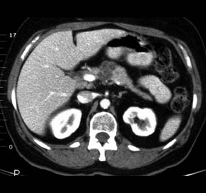
Cystic
Pancreatic Neoplasm

|
Mucinous cystadenomas are exocrine epithelial tumors of the
pancreas. Although uncommon, most occur in elderly women, and are
usually found in the tail or body of the pancreas. Half of these tumors
communicate with the pancreatic duct system. They are well-encapsulated
with a thick fibrous wall, and a multilocular internal structure. The
cysts are lined by mucinous columnar epithelium with papillary
proliferation, which show mild, moderate, or severe cellular atypia.
Indications of malignancy include irregular outline of the cyst wall
with tissue projecting into the lumen. Other signs of malignancy
include invasion of adjacent structures and metastases to the liver or
lymph nodes. In every
new case of a pancreatic pseudocyst, the diagnosis of cystic pancreatic
neoplasm should also be considered, especially if there are no known
causes.
Clinical symptoms develop insidiously, and laboratory studies provide
limited assistance. On US or CT, mucinous cystadenomas appear as
unilocular or multilocular neoplasms, with internal sepation, and
nodular tumor excrescences. Calcifications are occasionally present in
the periphery of the tumor. The tumor surface is smooth, with a thick
wall that may contain calcification. ERCP shows ductal encasement or
filling of an irregular cystic space (in cases of communicating
cystadenomas). Endoscopic US is the most sensitive technique for the
characterization of cystic neoplasms.
|
|
Radiologic Findings of Mucinous Cystadenomas |
 |
Multiple
non-enhancing cystic lesions, w/ lubulated external contour, showing enhancement of the cystic
wall structures after contrast injection. |
|
|


![]()
