GI Radiology > Pancreas > Trauma
Trauma
![]()
|
Traumatic injury to the pancreas is rare and difficult to diagnose.
Pancreatic injuries are present in 2% of closed abdominal trauma, far
behind injuries to the liver and spleen (47 and 44% respectively).
Vehicle accidents (involving abdominal impact against the steering
column) are the most common cause of pancreatic trauma. During violent
trauma, the pancreas is crushed against the vertebrae (L1). In order of
frequency, injuries to the pancreas involve the body, head, and tail.
These injuries are rarely isolated; 60% are duodenopancreatic lesions,
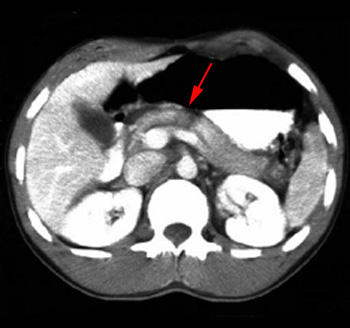
while 90% involve at least one other abdominal organ. During the initial evaluation, the symptoms of an isolated pancreatic lesion are often absent. Patients with early symptoms may report brief post-traumatic epigastric pain that has completely subsided. Pain may be absent for several days, even in the case of complete transaction with avulsion of the pancreatic duct. In isolated pancreatic lesions, the clinical manifestations may only appear with late complications, such as pseudocysts, abcesses, etc. Laboratory values are not very specific; the increase in serum and urine amylase levels can be delayed and non-specific. The presence of amylase in peritoneal lavage is indicative of pancreatic injury; its absence does not exclude injury, however. In view of the above, the history and mechanism of the impact often constitute the only criteria for suspecting the diagnosis. Common complications of pancreatic trauma include fistulas, pseudocysts, abcesses, and early pancreatitis. Fistulas are not always considered a complication since they may prevent the formation of pseudocysts (by providing a pathway for pancreatic enzyme drainage). Pseduoaneurysms of the pancreaticoduodenal arteries, secondary hemorrhage, and recurrent pancreatitis occur more rarely. Anatomic lesions are classified according to the damage to the parenchyma and ductal system. There are several classification systems; the table below lists one such system. |
||||||||||||||
|
Imaging:
ERCP may also assist in diagnosis of pancreatic trauma. It evaluates the integrity of the pancreatic duct, and identifies injuries that require surgical correction. ERCP is indicated in cases of high clinical suspicion but negative CT findings. |
|||||||||||||

