GI Radiology > Peritoneum > Air > Bowel Wall
Gas in the Bowel Wall (Pneumatosis Intestinalis)
![]()
|
Clinical Pneumatosis intestinalis can occur as a primary or secondary disorder. Primary pneumatosis intestinalis is less common (15%) and occurs when there is no other underlying respiratory or gastrointestinal abnormality. It is primarily a disease of older adults and is often asymptomatic. Secondary pneumatosis intestinalis is much more common (85%) and occurs int the setting of underlying bowel or pulmonary disease. It may be broken down into three subgroups: GI disease with bowel necrosis (i.e. necrotizing enterocolitis in infants, ischemic necrosis due to mesenteric vascular disease, strangulation, primary infection of the bowel wall); GI disease without bowel necrosis (i.e. pyloroduodenal peptic ulcers, bowel obstruction, IBD, connective tissue disease, endoscopy/colonoscopy, percutaneous jejunostomy tube, steroid therapy, leukemia, intestinal parasites, etc.); obstructive pulmonary disease (i.e. emphysema, bullous diseas, chronic bronchitis, asthma).
Radiological findings Primary pneumatosis intestinalis will appear as cystic gas in the colon on plain film and CT. Secondary pneumatosis intestinalis will appear as linear gas collections throughout the bowel wall.
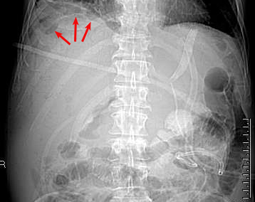
Plain film, from the patient with portal venous gas (arrows) on the previous page, demonstrates massive pneumatosis intestinalis as well.
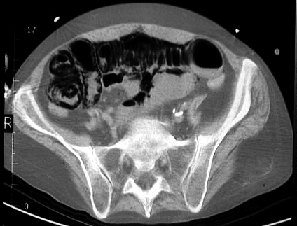
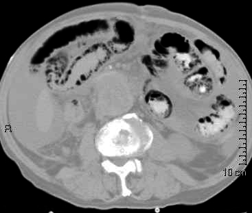
Two CT slices demonstrating massive pneumatosis intestinalis. Note the linear gas collections throughout the bowel wall.
|