GI Radiology > Peritoneum > Infectious > Peritonitis
Peritonitis
![]()
|
Clinical Peritonitis is simply inflammation of the peritoneum which may be caused by bacterial infection, granulomatous disease, or chemical irritation among other things. The process may be local (abcess) or generalized (peritonitis). It may occur as a primary process but more often occurs secondary to an intraperitoneal abcess or rupture of a hollow viscous.
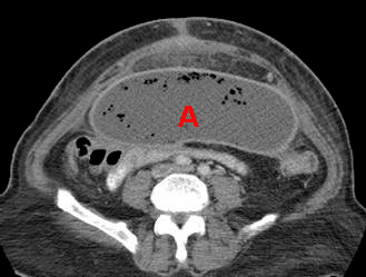
Radiological findings CT features of peritonitis include ascites in conjunction with peritoneal and mesenteric thickening. The CT appearance of an abcess is variable depending on the age of the abcess. An early abcess will approach soft tissue attenuation due to the presence of inflammatory cells and bacteria. As an abcess ages, it goes through liquefactive necrosis. If a definable wall (which may enhance) and a low attenuation center are present, than the abcess is mature. CT may also show thickening or obliteration of adjacent fat and displacement of adjacent structures. Gas bubbles within the fluid often indicate infection, but could also represent fistulous connection with bowel. The CT appearance of an abcess is similar to hematomas, bilomas, urinomas, necrotic tumors, and pseudocysts. MRI may be more useful in differentiating hematomas from abcesses, although it may miss small amounts of gas. Definitive diagnosis of a fluid filled mass is made by percutaneous aspiration.
This CT image shows an abcess (A) in the pelvis with a definable wall. The gas bubbles within the abcess raise the possiblity of an active infection, but they are relatively nonspecific. |