GI Radiology > Peritoneum > Quiz Answers
Answers to Peritoneum Quiz
|
1. In the supine position, the most dependent portion of the peritoneum in the upper abdomen where ascites will accumulate is: A. The pouch of Douglas B. The left subphrenic space C. The hepatorenal recess (Morrison's pouch) D. The lesser sac C: Morrison's pouch is the most dependent portion of the peritoneum in the upper abdomen. The pouch of Douglas is in the pelvis and is where fluid will accumulate in the upright position. |
|
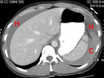
2. This CT from a trauma patient demonstrates:
A. Hemoperitoneum B. Splenic laceration C. Senitinel clot sign D. All of the above D: This CT shows a splenic laceration with consequent hemoperitoneum. The blood immediately adjacent to the spleen has a higher attenuation due to the fact that clotted blood is more dense, and this is known as the sentinel clot sign. |
|
3. The sentinel clot sign: A. Helps to localize the source of a hemopertioneum. B. Is due to the lower density of the clotted component of a hemoperitoneum. C. Is rarely seen. D. All of the above. A: The area of hemoperitoneum adjacent to a source of bleeding is most likely to clot first and may thus help localize the source of bleeding. The clotted component is MORE dense than the unclotted blood, not less dense. The sentinel clot sign is often seen. |
|
4. The most common cause of pneumoretroperitoneum is perforation of the first portion of the duodenum. True False FALSE: The first portion of the duodenum is intraperitoneal. The most common cause of pneumoretroperitoneum is perforation of the second, third, or fourth portions of the duodenum as these are all retroperitoneal. |
|
5. All of the following may be signs of free air in the abdomen EXCEPT: A. Double wall sign (Rigler's sign) B. Falciform ligament sign C. Football sign D. Ground glass sign D: The double wall sign describes the fact that both the inside and outside walls of bowel are visualized because they are outlined by air within the bowel and free air in the abdomen. The falciform ligament is visualized by free air in the abdomen. The football sign describes the shape of gas outlining the peritoneal cavity. The ground glass sign refers to the hazy appearance on plain film created by large amounts of fluid in the peritoneum. |
|
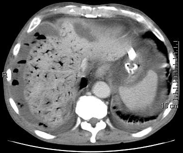
6. The following disease process may have been a result of:
A. Bowel wall necrosis secondary to mechanical obstruction. B. Infection of the bowel wall by gas producing organisms. C. Bowel wall necrosis secondary to mesentaric artery occlusion. D. Any of the above D: This CT shows a large amount of air in the portal venous system. This may result from any of the above mechanisms as they all allow gas to enter the bowel wall and travel to the portal venous system. There is also loculated gas and fluid in the peritoneum probably from bowel perforation. |