Gastrointestinal Radiology > Post-Test

Post-Test


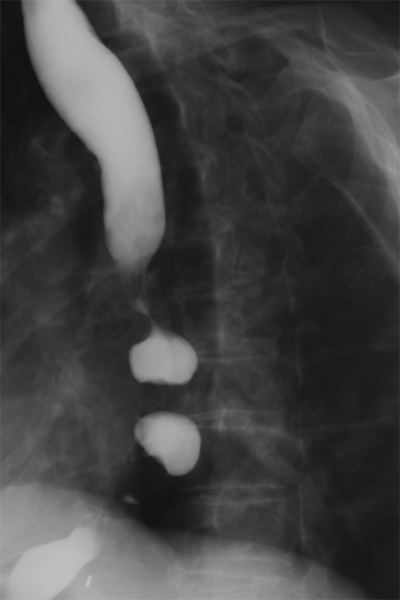
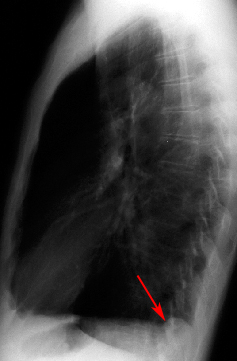
1. The appearance of the esophagus below is typical of what disease process:


A. Chagas' disease
B. Scleroderma
C. Diffuse esophageal spasms
D. Achalasia


2. The classic radiographic features of Barrett's esophagitis are high esophageal strictures or deep penetrating ulcers.
 

True
False


3. The majority of esophageal carcinomas are of the adenocarcinoma type.
 

True
False


4. Which of the following is NOT included in the differential diagnosis of enlarged gastric folds?
 

A. Zollinger-Ellison Syndrome
B. Linitis Plasitca
C. Lymphoma
D. Corrosive Gastritis
E. Menetrier's Disease


5. All of the following statements regarding gastric imaging are true EXCEPT:
 

A. Water soluble contrast should be used if a leak or perforation are suspected.
B. A double contrast upper GI series uses both oral and IV contrast.
C. A double contrast study provides improved mucosal visualization.
D. The imaging study of choice for diagnosing hypertrophic pyloric stenosis is ultrasonography.


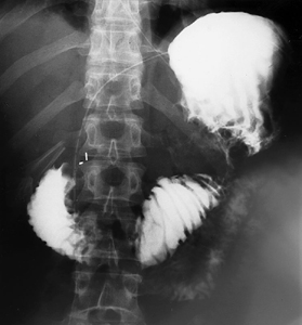
6. The image below is from a single contrast barium study in a 46 year old male following gastrojejunostomy. Based upon the findings below, what is the diagnosis?


A. Afferent limb syndrome
B. Bile reflux gastritis
C. Normal postoperative Billroth II
D. Gastric stump cancer
E. Anastomotic leak


7. What is the most common sequela of a Meckel's diverticulum?
 

A. Asymptomatic
B. GI bleeding
C. Obstruction
D. Perforation
E. Cancer


8. What structure is being imaged here?


A. Stomach
B. Ligament of Treitz
C. Terminal ileum
D. Rectum


9. What is the salient finding in the image in the above question?
 

A. Stricture
B. Fistula
C. Bowel dilatation
D. A and B
E. All of the above


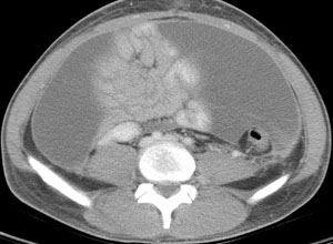
10. What is the abnormality in the image below?


A. Filling defect
B. Luminal dilatation
C. Stricture
D. Fistula


11. From where is the mass in the above image likely arising?
 

A. Bowel lumen
B. Bowel mucosa
C. Bowel wall
D. Extrinsically


12. The following image demonstrates a complication seen most often due to:


A. Appendicitis with a ruptured appendix
B. Metastatic carcinoid tumor
C. Metastatic mucinous adenocarcinoma


13.  What are the dimensions of the normal appendix as seen on ultrasound:
 

A. 7 cm x 2 cm
B. 7 mm x 2 mm
C. 7 in x 2 in
D. None of the Above


14.  Ischemic colitis is usually caused by an arterial occlusion.
 

True
False


15. Which of the following is malrotation NOT associated with?
 

A. Ulcerative colitis
B. Intussusception
C. Intestinal atresia


16.  Intussusception may be caused by any of the following EXCEPT:
 

A. Bacterial infection
B. Tumor
C. Meckel's diverticulum
D. Foreign body


17. Which of the following can be used to differentiate a paralytic ileus from a partial small bowel obstruction?
 

A. Distended loops on plain film
B. Dynamic air-fluid levels
C. Patient's symptoms


18.  Patients with suspected toxic megacolon should have a barium study to confirm diagnosis.
 

True
False


19.  Sigmoid volvulus is more common than cecal volvulus.
 

True
False


20. During what contrast phase is a hepatocellular carcinoma most likely to enhance?
 

A. Arterial phase
B. Portal venous phase
C. Equilibrium phase


21. All of the following are signs of advanced cirrhosis on imaging EXCEPT:
 

A. Liver surface nodularity
B. Contracted liver with ascites
C. Atrophy of the posterior segments (VI,VII) of the right lobe
D. Enlarged caudate lobe (I) and lateral segments (II,III) of the left lobe
E. Prominent umbilical vein
F. Irregular enhancement
G. All of the above are signs of advanced cirrhosis


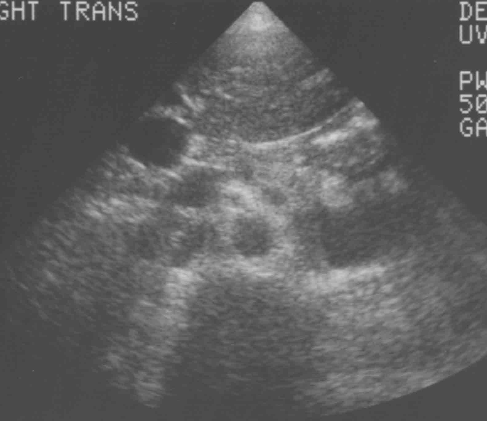
22.  The following image depicts a hepatic hemangioma.


True
False


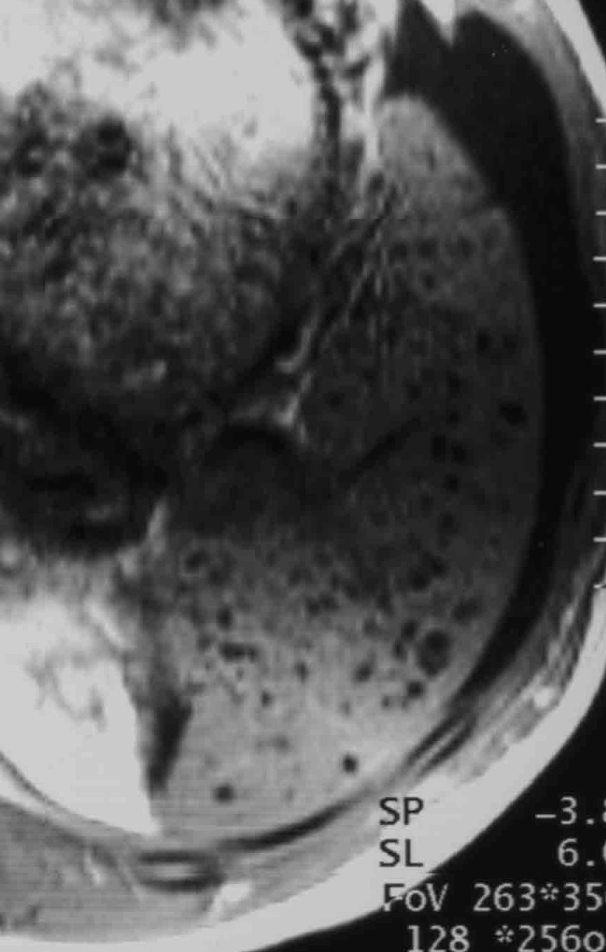
23.  The appearance of the liver below is due to:


A. CHF
B. Constrictive pericarditis
C. Restrictive cardiomyopathy
D. Any of the above


24.  The upper limit of normal for the dimensions of a gallbladder are:
 

A. 10 cm long, 3 cm wide
B. 15 cm long, 5 cm wide
C. 5 cm long, 3 cm wide
D. 20 cm long, 5 cm wide
E. 15 cm long, 10 cm wide


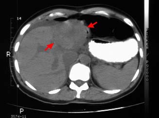
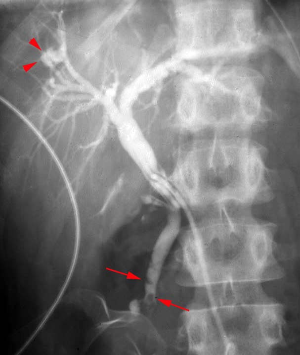
25. What condition does the following image most likely represent?


A. Ascending cholangitis
B. Biliary obstruction
C. Mirizzi syndrome


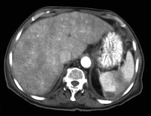
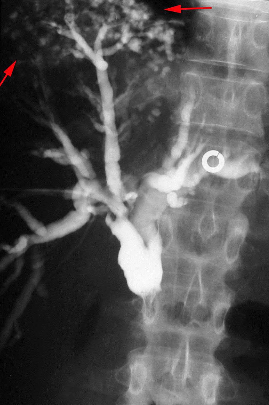
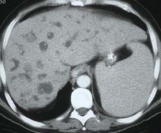
26. Caroli’s Disease is a type of choledochal cyst characterized by: (use the axial CT slice below)


A. Solitary fusiform extrahepatic cyst
B. Extrahepatic supraduodenal diverticulum
C. Intraduodenal diverticulum choledochocele
D. Multiple intrahepatic cysts


27.  All the following are radiographic signs of annular pancreas except:
 

A. Double bubble sign
B. Prenatal hydroamnios
C. Minimal distal bowel gas
D. Crescent sign


28.  The best imaging modality for acute pancreatitis is:
 

A. US
B. CT, no contrast
C. Contrast enhanced CT (oral & IV)
D. MRI


29.  All the following are CT signs of pancreatitis except:
 

A. Peripancreatic fluid collection & fat stranding
B. Focal non-enhancing regions of the pancreas on contrast-enhanced CT
C. Atrophic or enlarged pancreas with loss of normal internal structure
D. Duct dilatation and strictures
E. All of the above


30.  What condition could a patient have who presents with an MRI of the spleen seen on the right:
 

A. Splenic trauma
B. Abscess
C. Hepatitis C
D. Epidermoid cysts


31.  Which of the following is not part of the differential for the ultrasound seen on the right:
 

A. Metastatic disease
B. Abscesses
C. Posttraumatic cysts
D. All of the above are part of the differential


32. The sentinel clot sign:
 

A. Helps to localize the source of a hemopertioneum.
B. Is due to the lower density of the clotted component of a hemoperitoneum.
C. Is rarely seen.
D. All of the above.


33.  All of the following may be signs of free air in the abdomen EXCEPT:
 

A. Double wall sign (Rigler's sign)
B. Falciform ligament sign
C. Football sign
D. Ground glass sign


34.  Imaging of hernias is best achieved by:
 

A. Upper GI study then small bowel follow through.
B. CT, ultrasound, or herniography, since either technique is equally effective.
C. Depends on the clinical scenario, with each imaging modality providing different strengths.
D. CT, ultrasound, then MRI.


35. The following image demonstrates what type of hernia?


A. Morgagni hernia.
B. Petit hernia.
C. Grynfeltt hernia.
D. Bochdalek hernia.


36. The following image demonstrates what type of hernia? (R=rectus abdominus)


A. Bochdalek hernia.
B. Spigelian hernia.
C. Femoral hernia.
D. Direct inguinal hernia.


Your score is out of 36.

Do you want to see the answers?


© Copyright Rector and Visitors of the University of Virginia 2013