GI Radiology > Small Bowel > Neoplasms > Primary Adenocarcinoma
Neoplasms
![]()
Primary Adenocarcinoma |
ClinicalPrimary adenocarcinoma of
the small bowel is much less common than its dreaded colonic counterpart. It occurs
most commonly in the duodenum and jejunum. In fact, it is the most common malignancy of the duodenum.
Having small bowel adenocarcinoma increases the incidence of having other
primary malignancies by 8-fold. For example, 29% of patients also have
colorectal cancer. There is an increased
incidence in patients with celiac disease (duodenum, proximal jejunum) and
Crohn’s disease (ileum). Signs and symptoms nonspecific,
including abdominal pain, nausea, vomiting, weight loss, anemia.
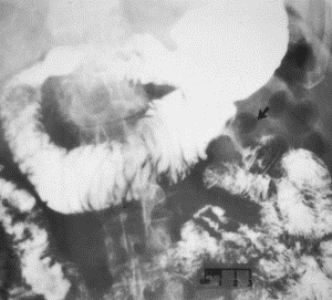
RadiologicalThe majority of primary adenocarcinomas occur within 25 cm of ligament
of Treitz. They can have a variable appearance radiographically, depending on
their location. In the duodenum (most common), the majority present as polypoid
filling defects, presumably from malignant transformation of adenomas. These
can ulcerate, leading to a “target” appearance on fluoroscopy. A small
minority of duodenal adenocarcinomas appear as infitrative masses with annular
luminal narrowing. Mesenteric small bowel adenocarcinoma, less common than duodenal adenocarcinoma, has a much higher propensity to present as an annular constricting lesion, with an “apple core” pattern and destroyed mucosa. Note, however, that 55% of small bowel annular lesions are metastases (as opposed to primary cancers). |
|
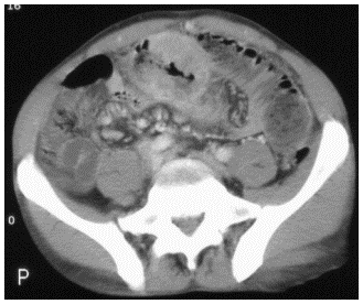
Primary adenocarcinoma. Fluoroscopy (left) demonstrates anannular luminal narrowing with shouldered margins (arrow). CT (right) demonstrates marked, irregular bowel wall thickening causing the “apple-core” appearance seen on fluoroscopy. |
![]()
![]()