Fluoroscopically Guided Balloon Dilation of GI Tract Strictures

Introduction
|
-
Strictures of the gastrointestinal tract usually
form as a complication of inflammation or carcinoma.
With the increasing number of surgical procedures involving
the gut, the number of gastrointestinal strictures complicating
the formation of a surgical anastomosis has also risen.
Whatever the cause, the strictures are usually persistent,
and they may produce significant clinical symptoms.
-
The standard nonoperative management of
gastrointestinal strictures has been with graded dilators of
progressively larger diameter.
As strictures are most common in the esophagus, treatment
has been traditionally focused on this portion of the gut.
For many years, bougienage has been the standard treatment
to dilate the strictures. The
more recent method of stricture dilation involves the use of
balloon catheters with fluoroscopic guidance.
This technical advance became feasible only after the
introduction of a double-lumen catheter with a low-compliance
balloon, originally intended for transluminal angioplasty.
This device permits generation of high pressure in the
balloon while maintaining a fixed balloon diameter.
-
Balloon dilation offers a number of advantages
over bougienage. The
most important is that the balloon remains in a stationary
position during inflation and applies only radially directed
forces to the gut wall. Forces
are maximal at the narrowest point, the stricture itself.
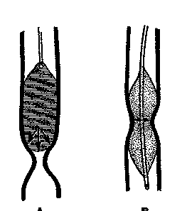
In contrast, a bougie must exert considerable longitudinal
force on the gut wall to generate an effective radial dilating
force on the stricture. This
longitudinal shearing force increases the risk of rupture and
mucosal injury which can lead to recurrence of the stenosis (Fig.
1).

Fig.
1.
Dilation of esophageal stricture with bougie or balloon.
(A)
The bougie is lowered down onto the stricture. The
dilating forces are chiefly
longitudinal on the gut wall (arrows).
(B)
The balloon is positioned across the
stricture. During inflation, the dilating forces (arrows)
are only radially directed to the gut wall resulting in more
effective stretching of the stricture. (From
Shaffer & de Lange, ref. 7.
Reproduced by permission.)
-
The adverse effect of the longitudinal shearing
force is demonstrated by the post-dilation relapse-free interval,
which averages about six times longer after balloon dilation than
after bougienage in the treatment of esophageal strictures.
Another advantage of using a balloon is that dilation
pressure can be monitored and controlled with an in-line pressure
gauge. Also, balloon
dilation may be more comfortable for the patient than bougienage
because it usually requires only a single passage of a small
catheter with a collapsed balloon, whereas bougienage requires
multiple exchanges for progressively larger and larger dilators.
-
Fluoroscopically guided dilation has advantages
over blindly performed or endoscopically controlled procedures.
When endoscopy is used to guide a dilating bougie or
balloon, only the gut proximal to the stricture is visualized once
the dilator enters the stricture. Beyond that point, the instrument is passed without visual
control, and perforation by the tip of the dilator can occur.
Fluoroscopy allows visualization of the stricture as well
as the gut proximal and distal to it.
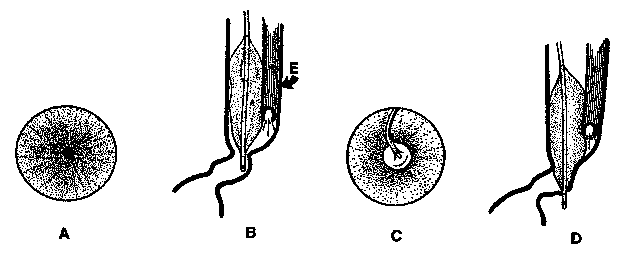
It also permits visual control of the entire balloon
catheter during its placement and inflation (Fig. 2).

Fig.
2. Endoscopically
guided balloon dilation of strictures in tortuous gut.
(A)
View through the endoscope positioned at the stricture in
the gastrointestinal tract shows only the orifice of the
stricture.
(B)
The tortuosity of the gut more distally is not seen
endoscopically. When the balloon is advanced, the orifice becomes obscured. E
= endoscope.
(C)
View through the endoscope with the balloon in the
stricture. Only the
proximal portion of the balloon
is visible to the examiner and there is no control of the tip of
the catheter.
(D)
When the balloon catheter is advanced, a perforation can
easily occur and is likely to go unnoticed as the tip of the
catheter is not visible to the examiner.
(From Shaffer & de Lange, ref. 7.
Reproduced by permission.)
|
|
|


![]()
![]()