-
Again,
place the patient in the sitting position on the Brunswick Chair mounted
on the footrest of the upright fluoroscopic table. The entire
examination is performed with the patient seated in the right lateral
projection.
-
Position the fluoroscope so that the field of view includes the
sacrum posteriorly, the symphysis pubis anteriorly, and an area about
two inches below the skin surface of the perineum (to allow for downward
descent during evacuation). Also include the commode's centimeter marker
within the field of view to permit later measurement corrections for
magnification.
-
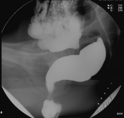
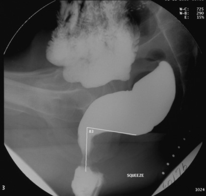
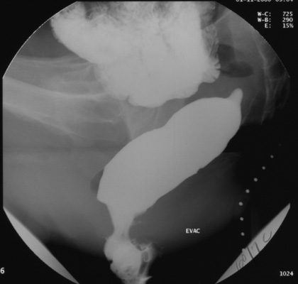
Take digital images during each of the following maneuvers:
-
in the resting state
-
with maximal voluntary contraction of
the sphincter and pelvic floor muscles ("squeeze")
-
intermittently
during defecation
-
Videofluorography is also done while
the patient performs the following maneuvers: resting state, squeezing and finally, rectal evacuation. In the latter phase of the
study, key events can also be recorded with intermittent digital imaging.
|