GI Radiology > Spleen > Others > Gamna-Gandy Nodules
Iron Deposition Disorders: Gamna-Gandy Nodules
![]()
-
Gamna-Gandy bodies are hemosiderotic nodules secondary to perifollicular and trabecular hemorrhage in the spleen. The hemorrhagic infarcts often result in any condition resulting in congestive splenomegaly, due to deposition of hemosiderin and calcium. MRI magnetic imaging (T1W & T2W sequences) are highly sensitive.

Hx: 22 yo with cystic fibrosis, liver dz, pancreatic insufficiency
-
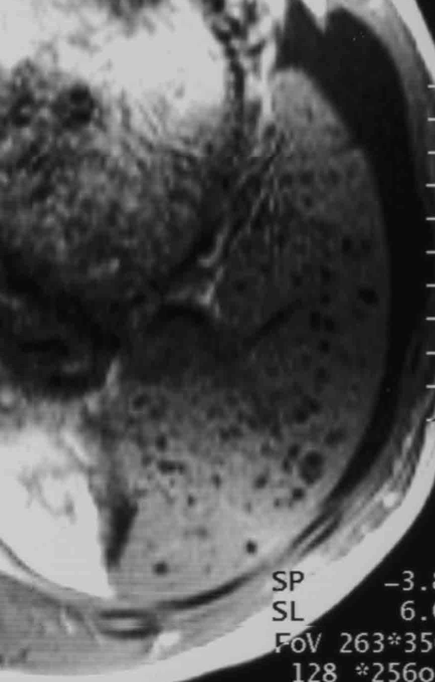
The above patient demonstrates multiple Gamna-Gandy nodules, appearing as numerous low signal defects on FLASH images and as hyperechoic spots on ultrasound. In the images shown the sizes range from 3 - 8 mm. The patient shows signs of portal hypertension with massive splenomegaly.

Hx: Hep C, EtOH hepatic cirrhosis with portal hypertension
-
On the MRI above, you can see many hypointense lesions in the spleen that do not enhance even after administration of gadolinium. These lesions, known as Gamna-Gandy bodies, represent sites of hemosiderin deposition in the spleen. There are two possible diagoses to consider with this presentation on MRI:
- Hemosiderosis
- Increased deposition of iron without organ damage. This iron deposition usually occurs in the spleen, lymph nodes, and liver
- Hemochromatosis
- Increased iron in the liver and other organs with organ damage. Two seperate categories include:
- primary - idiopathic or genetic cause
- secondary - chronic anemias with multiple blood transfusions, ingestion of excessive iron, cirrhosis
