GI Radiology > Small Bowel > Functional Abnormalities > Sprue
Functional Abnormalities
![]()
Sprue |
ClinicalCeliac disease (non-tropical sprue, gluten-sensitive enteropathy) is an autoimmune disorder in which dietary gluten (wheat, barley, rye) damages small bowel mucosa, causing malabsorption. Histologically, this presents as villous atrophy, crypt hyperplasia, and chronic inflammation. Symptoms include diarrhea, steatorrhea, abdominal pain, and distention. Signs of malabsorption accompany these symptoms, including anemia, bleeding, neuropathy, glossitis, weight loss, and malaise. Diagosis is usually made through iestinal biopsy. Treatment is comprised of a gluten-free diet. Patients with sprue demonstrate a 10-15% incidence of GI malignancy, 50% of which are lymphoma. Tropical sprue occurs in
tropical regions and is radiographically similar to celiac disease. It is associated
with severe B12 and folate deficiencies, causing megaloblastic anemia. It is not
associated with gluten sensitivity and is treated with folic acid supplements
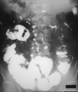
and antibiotics. RadiologicalCeliac disease is the prototype for all malabsorption syndromes. Fluoroscopy is the most sensitive imaging study for evaluation. The small bowel lumen is often dilated. Folds are normal thickness, but fold patterns are altered. There is a decreased number of folds in the jejunum (<3/inch) and an increased number in the ileum (“jejunalization”). In addition, there is a “wet” or “hypersecretion” pattern, caused by increased fluid in the bowel lumen from malabsorption. This “wet” pattern is comprised of barium dilution, indistinct folds, and flocculation. Another interesting phenomenon associated with celiac disease is transient intussusceptions, which are non-obstructive and painless. |
|
Small bowel follow-through in a patient with celiac
sprue. Initial imaging (left) demonstrates mild dilatation and “jejunalization”
of the ileum. Subsequent imaging at 30 minutes (right) demonstrates a rapid
transit time, with barium dilution and flocculations. |
![]()
![]()