GI Radiology > Small Bowel > Structural Abnormalities
Structural Abnormalities
![]()
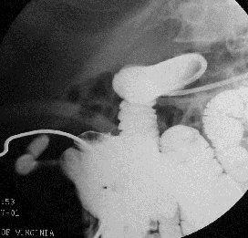
Meckel’s Diverticulum |
|
Meckel’s diverticulum is
the failed obliteration of intestinal end of omphalomesenteric duct. It is a true diverticulum, containg
all three muscular layers of the bowel wall. It occurs in the distal ileum,
usually within 100cm of the ileocecal valve. It is common, present 2-3% of
autopsies. The majority of Meckel’s diverticula are asymptomatic, and 50%
contain heterotopic mucosa (usually gastric, or less commonly pancreatic).
Those that do present clinically have variable presentations. The most common
presentation in adults is GI bleeding, a result of ectopic gastric mucosa.
Children more often present with small bowel obstruction, as the diverticulum
acts as a lead point for intussusception. Rarer complications include
diverticulitis (“pseudoappendicitis”) or perforation. RadiologicalPlain films are often
normal, but demonstrate distal SBO or enteroliths. Generally, plain films do
not play a major role in the diagnosis of Meckel’s. Fluoroscopy demonstrates
the diverticulum as a contrast-filled outpouching from the distal ileum.
Rugae may be seen as a result of ectopic gastric mucosa, and and
complications, such as intussusception or ulceration, may be identified. Nuclear medicine can aid
in the diagnosis of Meckel’sdiverticulum, but only if it contains gastric
mucosa. A “Meckel’s scan” (technetium-99m-pertechnetate) selectively
localizes to gastric mucosa, with a positive study showing abnormal activity
in the lower abdomen. This test is particularly effective at identifying
Meckel’s diverticula as the cause of GI bleeding, because a bleeding Meckel’s
diverticulum invariably contains ectopic gastric mucosa. |
|
Meckel’s diverticulum. Enteroclysis demonstrates a saccular outpouching. Meckel's scan (Tc-99m-labeled RBCs) shows abnormal uptake in the lower abdomen. Increased uptake is seen in diverticula that contain ectopic gastric mucosa. |
![]()
![]()