GI Radiology > Small Bowel > Structural Abnormalities
Structural Abnormalities
![]()
Intussusception |
|
Intussusception is the
telescoping of bowel to form an inner loop (intussusceptum) and an outer loop
(intussuscipiens). In children 95% of intussusceptions are idiopathic, with
lymphoid hyperplasia or hypertrophy postulated as possible causes. In adults,
intussusception is usually caused by a neoplasm or other mass acting as a
lead point. Intussusception often
presents as small bowel obstruction, with cramping abdominal pain, vomiting,
and blood in the stool (currant-jelly stool). With prompt reduction, bowel
necrosis can be avoided. Prolonged intussusception may result in bowel
necrosis and perforation. Surgery is recommended when radiological reduction
is unsuccessful. Radiological Plain film may be normal. Often
there is an absence of gas in right colon (especially in children). Bowel dilatation
signifies obstruction, and intraperitoneal free air signifies perforation. Fluoroscopy
demonstrates a coil-spring appearance, with barium trapped between
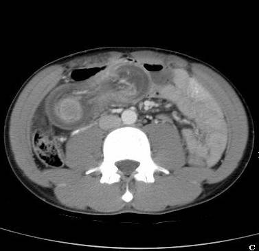
intussusceptum and intussuscipiens. CT scan shows a “bull’s-eye,” which
represents the intussusceptum and mesentery surrounded by intussuscipiens. Ultrasound
is effective at identifying itussusception. Viewed in the transverse plane, intussusception
is seen as a swirled pattern of alternating hyperechogenicity and
hypoechogenicity. The longitudinal view demonstrates a “sandwich” appearance
of the alternating loops of bowel.
Intussusception. CT demonstrates edematous bowel wall with a target appearance. The intussusceptum forms the inner part of the bull’s eye, while the intussuscipiens forms the outer layer. |
|
|
![]()
![]()