GI Radiology > Appendix > Anatomy
Anatomy of the Appendix
![]()
|
The appendix is a small, worm-shaped blind tube, about 3 in. (7.6 cm) long and 1/4 in. to 1 in. (.64*2.54 cm) thick, projecting from the base of the cecum on the right side of the lower abdominal cavity. It has no known function in humans today and is considered a vestigial remnant of some previous organ or structure, having a digestive function, that became unnecessary to people in their evolutionary progress.
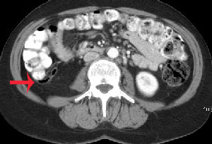
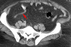
The appendix can usually be identified on an abdominal CT. This can be accomplished by identifying the ascending colon and following it caudally to the cecum. One should then look medially for the junction of the terminal ileum with the cecum and locate the ileocecal valve. The appendix is usually found caudal to the ileocecal valve at the base of the cecum. Look for a small, blind ending tubular structure projecting from the cecal base.
If you are unable to identify the appendix in this
manner, you can try to give the patient more oral contrast in hopes of filling the appendix or repositioning the patient on their side left side down and reimaging.
Abdominal CT scans from two different individuals demonstrating normal appendices. In the top image note the air filled appendix projecting from the base of the cecum. In the bottom image note the contrast filled, blind ending, tubular appendix projecting from the cecal base.
|
|
|


