Secondary neoplasms may cause biliary obstruction. Possible sources include (top to bottom):
Hepatocellular carcinoma.
Pancreatic carcinoma.
Ampullary tumor: occur at the ampulla of Vater and originate from the bile duct, pancreas, or duodenum.
Periampullary duodenal tumors.
Radiographic findings:
Hepatocellular carcinoma
Radiographic findings can be found in the liver section.
Pancreatic carcinoma
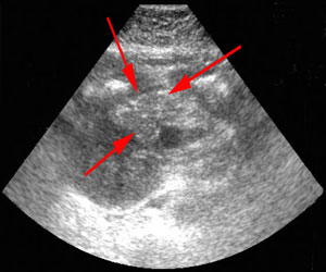
US: in 95% of cases, a low echogenicity mass is shown causing bile duct obstruction.
Note hypoechoic mass ahead of pancreas (arrow).
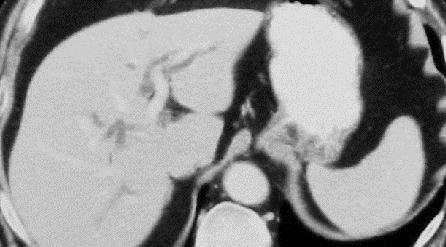
On CT, the following will likely be apparent: bile duct and pancreatic duct dilation (double duct sign); abrupt, focal, irregular stricture of the common biliary duct; bile duct encasement by direct extension or metastasis to peribiliary nodes.
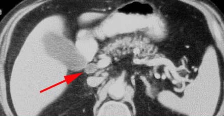
CT shows pancreatic head mass (arrow).
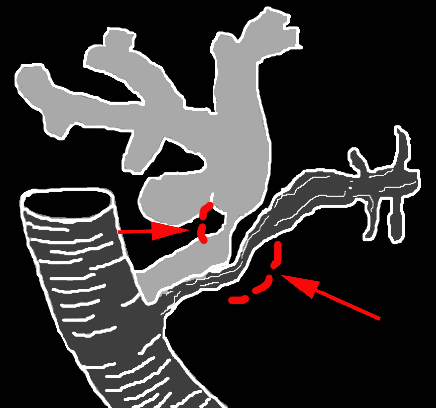
The diagram below shows a typical appearance of pancreatic carcinoma on ERCP.
Ampullary tumor
Biliary dilation is seen proximal to the tumor.
Figures: Choledocholithiasis and ampullary carcinoma. (A) ERCP shows stones floating in the lumen of the common bile duct and tumor protruding from the wall of the distal common bile duct (arrow).
A
(B) CT shows dilated intrahepatic bile ducts due to common bile duct obstruction. (C) CT shows tumor surrounded by contrast material in dilated distal common bile duct (arrow).