- Pathogenesis:
- Causes of cirrhosis include: a) alcohol, b)
alpha1-antitrypsin, c) postnecrotic (hepatitis), d) metabolic disease:
Wilson, hemochromatosis, glycogen storage disease, e) PBC, PSC, f)
congestive heart failure
- The most common cause in North America
is alcohol abuse.
- Cirrhosis pathology consists of
hepatocyte necrosis, fibrosis, and nodular regeneration.
- Cirrhosis increases risk of developing
hepatocellular carcinoma. The risk of malignancy is also increased with
several other causes of liver failure, including hemochromatosis,
Wilson's disease, aflatoxin, and chronic active hepatitis.
- In cirrhotic liver, a regenerating
nodule may resemble HCC.
- Radiographic findings:
- Signs of advanced cirrhosis on imaging:
- Liver surface nodularity
- Contracted liver with ascites
- Atrophy
of the posterior segments (VI, VII) of the right lobe
- Enlarged caudate lobe (I) and lateral
segments (II, III) of the left lobe
- Prominent umbilical vein
- Irregular enhancement
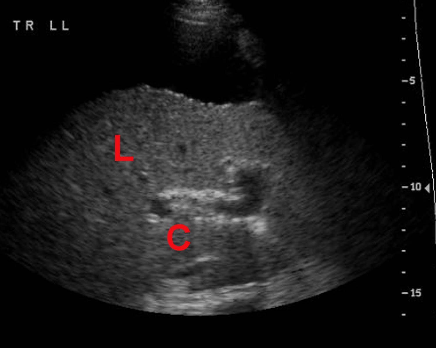
- U/S: advanced cirrhotic liver appears to
be nodular, irregular, and contracted with relatively enlarged caudate
lobe (C) and lateral segment (L) of the left lobe. Fatty infiltration and
fibrosis give a coarse echotexture of the liver parenchyma.
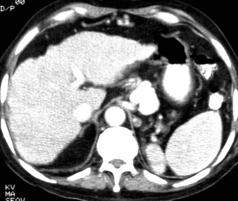
- Noncontrast CT: depending on the extent
of fatty infiltration, fibrosis, and regenerating nodules, the hepatic
parenchyma may have either homogeneous or heterogeneous decreased
attenuation.
- Contrast CT: areas of fibrosis and
regeneration may become isodense to parenchyma. The surface of the liver may be very nodular in cirrhosis.
- T1-weighted MRI: looks normal with
slightly heterogeneous signal intensity as with T2 except that areas of
fibrosis may be of low signal.
- T2-weighted MRI: looks normal with
slightly heterogeneous signal intensity as with T1 except that areas of
fibrosis may be of high signal.
|