GI Radiology > Small Bowel > Neoplasms > Lymphoma
Neoplasms
![]()
Lymphoma |
||
|
Lymphoma, because of its ability to
affect lymphoid tissue throughout the body, as well as its ability to “do
anything and go anywhere,” is difficult to exclude from any differential
diagnosis. The answer to the question, “Could this be lymphoma?” is almost
invariably, “Yes.” This holds
true for the small bowel. The small bowel is the most-often affected
intestinal site of lymphoma, usually representing secondary involvement of
non-Hodgkin’s lymphoma. It most often affects the distal ileum, because if
its predominance of lymphoid tissue (remember Peyer’s patches?). There is an
increased risk in patients with celiac disease or immunodifficiency. As
expected by its spectrum of appearances, it has an extremely variable
clinical presentation.
On imaging studies,
lymphoma has the “squirrelly” ability take on just about any radiographic
appearance. Its grocery-list
appearance includes fold thickening and effacement, luminal narrowing, aneurysmal
bowel wall dilatation, diffuse nodularity, extrinsic compression from
mesenteric masses, solitary or multiple filling defects, ulceration, intussusception,
to name a few. (There is no need to memorize this list, as you will notice it
pretty much encompasses the entire gamut of small bowel disease processes.) If your brain is
running out of room for information (like mine), you may find it easier to
just add lymphoma to all of your differential diagnosis lists. |
||
|
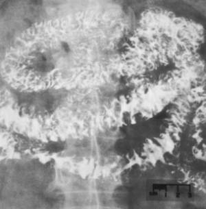
Diffuse, irregular fold thickening in lymphocytic lymphoma. |
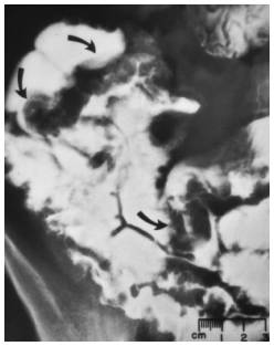
Multiple filling defects in lymphocytic lymphoma. The arrows point out the defects. Note the “target” appearance of the lesions, consistent with malignancy |
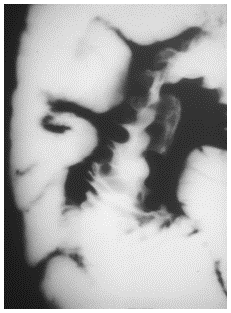
Ulceration in histiocytic lymphoma. Note also the luminal narrowing and fold thickening. |
![]()
![]()