GI Radiology > Small Bowel > Neoplasms > Carcinoid
Neoplasms
![]()
Carcinoid |
RadiologicalCT
is essential in the evaluation of carcinoid tumors. While less sensitive than
fluoroscopy at evaluating the mucosal surface, it is far more effective at
evaluating the extent of bowel wall involvement and size of extraluminal
mass. In addition,
extraintestinal spread into the adjacent soft tissues can be seen, as well as
metastatic spread to the mesentery, lymph nodes, and liver. As discussed
before, mesenteric metastases are often larger than the primary tumor,
appearing as a stellate, calcified mass with associated desmoplastic
reaction. Liver metastases are usually hypervascular, most prominent in the
early arterial phase of contrast enhancement. |
|
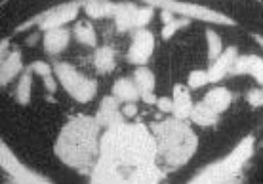
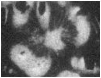
Carcinoid tumor. CT (left) demonstrates a large hyperdense mesenteric mass. Magnification of the mass (right) clearly shows the stellate calcifications that are often seen in advanced carcinoid tumors. |
![]()
![]()