GI Radiology > Small Bowel > Neoplasms > Carcinoid
Neoplasms
![]()
Carcinoid |
RadiologicalBecause signs and symptoms
are often absent or nonspecific early in the disease course), imaging plays
an important role is disease identification. Early (before muscular invasion
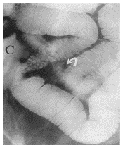
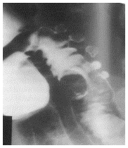
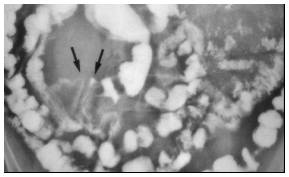
and desmoplastic metastases) manifestation is that of a solitary polypoid
nodule projecting into the bowel lumen, or less frequently, multiple nodules.
Because of its sensitivity
in evaluating the mucosal surface, fluoroscopy is optimal for demonstrating
small carcinoid tumors. Fluoroscopically, these nodules are seen as smooth,
rounded luminal filling defects. As the mass enlarges, hemorrhage and
necrosis can occur. If growth
occurs into bowel lumen (endoenteric), this can result in ulceration or
obstruction. Rarely, carcinoid can be the lead-point in intussusception. If
growth occurs into the soft tissues adjacent to the bowel wall (exoenteric
growth), it can cause separation and tethering of bowel loops and a
desmoplastic (fibrotic) reaction.
(click on images to enlarge)
|
![]()
![]()