Small Bowel Follow-Through (cont.)

The Radiologic Procedure
|
- The patient is placed in the prone
position for overhead radiographs.
- In this position, the center of the
abdomen is compressed, making the entire abdomen more uniform in
thickness, thus permitting more uniform x-ray penetration through the
small bowel.
- Another advantage to the prone position is that there is
better separation and less overlapping of small bowel loops.
- Additionally,
loops of ileum tend to migrate cephalad and become less compacted in the
pelvis when the patient is lying on his/her abdomen.
- In the interval between overhead radiographs, the patient
is generally allowed to walk about or sit in a dressing booth outside the
x-ray room.
- A debilitated patient can be left on the x-ray table between
radiographs or can be moved to a stretcher outside the room if it is needed for
other examinations.
- The patient who is unable to sit or stand should be
placed in the right-side-down position to encourage gastric
emptying-and then rolled periodically from one side to the other to better
distribute the barium and speed its transit by using the assistance of
gravity.
- Overhead radiographs are made on 14 x 17 inch (35 x
43 cm) format.
- The interval between exposures depends
on the speed of barium transit through the small bowel. Routinely, we
obtain films at 15-minute intervals for the first hour and at 30-minute
intervals thereafter.
- Each overhead radiograph should be carefully
examined as soon as it is processed. Any suspected abnormality should be
evaluated with fluoroscopy and compression spot images.
- Many authorities recommend, even in the
absence of abnormalities on the overhead films, that periodic
fluoroscopic inspections and compression spot imaging be performed.
- In any case, after the barium has
reached the right side of the colon, compression spot images of the
terminal ileum are routinely obtained. The terminal ileum is the most common location of small bowel pathology. Representative compression spot images of small bowel in all four quadrants of the abdomen are obtained to demonstrate that the entire small bowel was evaluated fluoroscopically.

|
|
|
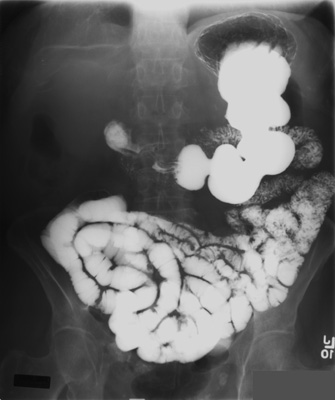
Figure: Normal Small Bowel


![]()
![]()